Lecture 7: Electron Optics, Aberrations, and Probe Formation in SEM
Автор: Science Tech Engineering and Math with Dr. Tiwari
Загружено: 2026-02-13
Просмотров: 7
Описание:
In this lecture, we move from electron generation to electron control. After understanding how high-quality electron beams are produced using thermionic and field emission sources, we now focus on how that beam is preserved, shaped, and focused as it travels through the microscope column.
The central topic of this lecture is electron optics — the system of magnetic lenses, apertures, and deflectors that determine probe size, beam current, and ultimately spatial resolution in scanning electron microscopy.
We begin by introducing the fundamental difference between light optics and electron optics. Because electrons carry charge, their trajectories can be controlled using electric and magnetic fields rather than glass lenses. While electrostatic lenses are conceptually simple, they suffer from strong chromatic aberration and are therefore rarely used for high-resolution SEM focusing. Modern SEMs rely primarily on magnetic lenses based on solenoid coils and pole pieces.
We then develop a physical understanding of magnetic lens operation:
• Lorentz force and spiral electron motion
• Lens current control of focal length
• Effects of accelerating voltage and working distance
• Role of condenser and objective lenses in probe formation
From there, we examine the fundamental limitations of real lenses:
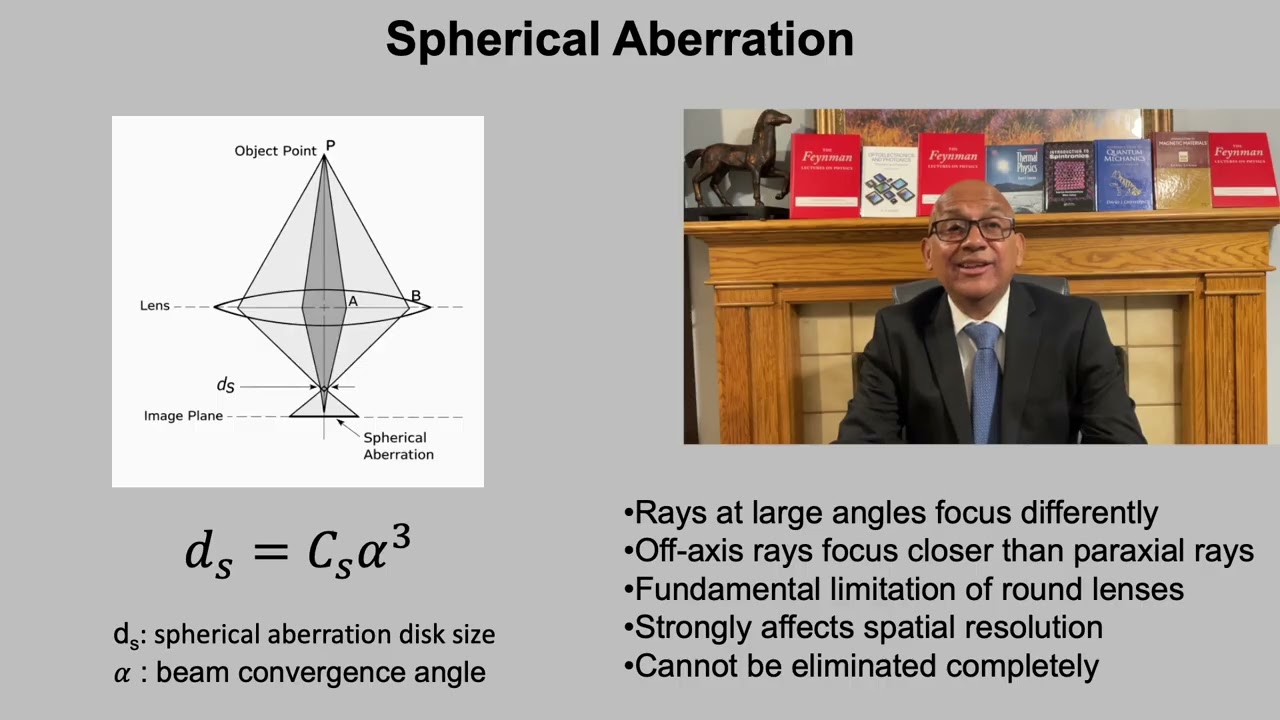
• Spherical aberration (cubic dependence on convergence angle)
• Diffraction (wave nature of electrons and Airy disk formation)
• Chromatic aberration (energy spread effects)
We show how these competing effects lead to an optimum aperture angle, where spherical aberration and diffraction are balanced. This establishes a fundamental resolution limit in conventional round electromagnetic lenses.
The lecture then shifts to practical operator controls and how they influence probe formation:
• Objective aperture size (controls maximum ray angle)
• Working distance (controls lens geometry and resolution vs depth of field trade-off)
• Condenser lens strength (controls beam current and convergence angle)
We introduce the deflector system and raster scanning, emphasizing that SEM magnification is determined by scan amplitude rather than lens magnification. This leads naturally to the concept of pixel size and sampling.
A major conceptual development in this lecture is the distinction between:
• Probe size (set by electron optics)
• Pixel size (set by scanning and sampling)
• Interaction volume (set by electron–specimen physics)
We demonstrate that SEM resolution depends on the balance between probe size and specimen pixel size. Magnification alone does not improve resolution. If the probe is too large, the image blurs. If the pixel size is too large, fine features are averaged out. True resolution requires coordinated optimization of optics and sampling.
By the end of this lecture, students understand that SEM imaging is not simply about “focusing the beam,” but about managing multiple competing limitations — aberrations, diffraction, chromatic effects, sampling density, and beam–specimen interaction.
In the next lecture, we will complete this framework by examining the third fundamental factor that limits SEM resolution: the electron–sample interaction volume and signal generation depth.
📌 Course: MSE 3011 – Structural Analysis of Materials
🎓 Instructor: Prof. Ashutosh Tiwari, University of Utah
#MaterialsScience
#StructuralAnalysis
#ElectronMicroscopy
#SEM
#ElectronOptics
#MagneticLenses
#Aberrations
#ProbeFormation
#Nanomaterials
#MaterialsCharacterization
#EngineeringEducation
#UniversityOfUtah
#MSE3011
Повторяем попытку...
Доступные форматы для скачивания:
Скачать видео
-
Информация по загрузке: