Knee Ultrasound: Articular Cartilage Pathologies (Chondromalacia, OCD, Chondrocalcinosis)
Автор: Dobry ortopeda Warszawa
Загружено: 2026-02-13
Просмотров: 28
Описание:
This video demonstrates ultrasound evaluation of articular cartilage pathology in the knee joint, including:
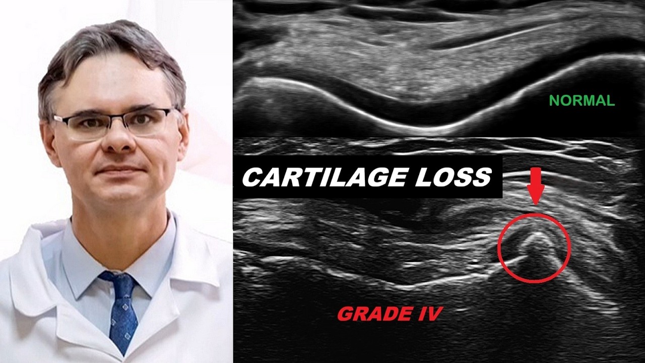
Chondromalacia (Grades I to IV)
Chondrocalcinosis
Osteochondritis dissecans (OCD)
Advanced degenerative cartilage loss
Subchondral bone irregularity
Indirect ultrasound signs of osteoarthritis
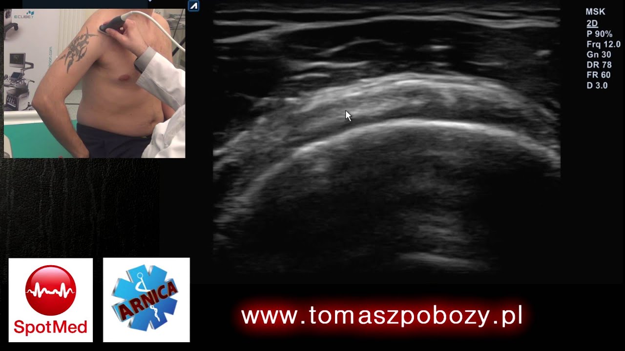
The examination is performed using high-frequency musculoskeletal ultrasound in routine orthopedic clinical practice.
Ultrasound allows dynamic, real-time assessment of femoral condylar cartilage, trochlear groove cartilage, and weight-bearing surfaces of the knee. Proper probe alignment and recognition of anisotropy artifacts are essential for accurate interpretation.
What is shown in this video?
Normal cartilage appearance in adult and pediatric knees
Cartilage thickness assessment, approximately 2 millimeters in adults
Grade I chondromalacia - softening, thickening, increased echogenicity
Grade II - partial-thickness defects less than 50 percent
Grade III - cartilage thinning greater than 50 percent
Grade IV - complete cartilage loss with subchondral irregularity
Osteophyte formation in advanced osteoarthritis
Chondrocalcinosis - intracartilaginous calcifications
Osteochondritis dissecans (OCD) in longitudinal and transverse planes
This presentation emphasizes ultrasound pattern recognition, clinical correlation, and practical diagnostic pitfalls.
Osteochondritis Dissecans (OCD) - Scientific Background
OCD predominantly affects the medial femoral condyle and is frequently observed in young athletes. Early detection is essential to prevent progression to osteoarthritis.
Related peer-reviewed publications:
Sport-Specific Risks of Osteochondritis Dissecans Across Athletic Disciplines: A Narrative Review
Pobozy T. et al., Healthcare, 2025
https://doi.org/10.3390/healthcare131...
Understanding Osteochondritis Dissecans: A Narrative Review of the Disease Commonly Affecting Children and Adolescents
Konarski W., Pobozy T. et al., Children (Basel), 2024
https://doi.org/10.3390/children11040498
A Comparative Analysis of Osteochondritis Dissecans and Avascular Necrosis: A Comprehensive Review
Konarski W., Pobozy T. et al., Journal of Clinical Medicine, 2024
https://doi.org/10.3390/jcm13010287
Basic Differences and Most Common Findings in Musculoskeletal Ultrasound in Children
Pobozy T. et al., Healthcare, 2022
https://doi.org/10.3390/healthcare101...
These publications provide deeper insight into epidemiology, sport-specific risk factors, diagnosis, and treatment strategies in OCD and related cartilage disorders.
Who is this video for?
Orthopedic surgeons
Sports medicine physicians
Musculoskeletal radiologists
Physiotherapists
Medical students
MSK ultrasound trainees
This material is educational and does not replace clinical examination, MRI, or radiographic imaging when indicated.
Equipment
The examination was performed using:
Alpinion eCube XC90 Elite
High-frequency single crystal linear transducer (3 to 19 MHz)
High-resolution imaging allows detailed assessment of cartilage morphology and subchondral bone interface.
Further MSK Ultrasound Content
Explore the full MSK Ultrasound Clinical Orthopedic Cases playlist for:
Meniscal pathology
Ligament injuries
Dynamic knee examination
Baker's cyst
Pediatric MSK ultrasound
Degenerative joint disease
Accurate cartilage evaluation requires experience, optimal probe positioning, and integration of clinical findings.
Thank you for watching.
Tomasz Pobozy, MD, PhD
Orthopedic Surgeon
Musculoskeletal Ultrasound
#KneeUltrasound
#Chondromalacia
#OsteochondritisDissecans
#MSKultrasound
#CartilageDamage
00:00 Normal Articular Cartilage - Introduction
00:18 Normal Cartilage - Trochlear Groove Transverse
01:07 Normal Cartilage - Medial and Lateral Femoral Condyles Anterior
01:29 Normal Cartilage - Medial Femoral Condyle Posterior
01:51 Normal Cartilage - Lateral Femoral Condyle Posterior
02:11 Thick but Normal Cartilage in Children - Trochlear Groove
02:32 Thick but Normal Cartilage in Children - Femoral Condyles
02:51 Chondromalacia Grade I to II - Trochlear Groove
03:18 Chondromalacia Grade II - Surface Irregularity
03:35 Probe Angle Artifact vs True Cartilage Damage
04:15 Chondromalacia Grade II to III vs Grade IV - Comparative View
04:46 Advanced Chondromalacia - Grade III to IV
05:00 Extensive Grade IV Cartilage Loss and Osteophyte
05:20 Chondrocalcinosis - Intracartilaginous Calcifications
05:38 Indirect Signs of Advanced Degeneration - Medial Compartment
06:17 Osteochondritis Dissecans OCD - Longitudinal View
06:49 Osteochondritis Dissecans OCD - Transverse View
06:59 Diagnostic Pitfalls in OCD
07:15 Clinical Interpretation and Final Remarks
Повторяем попытку...
Доступные форматы для скачивания:
Скачать видео
-
Информация по загрузке: