USMLE Renal 1: Renal Anatomy and Urinary Incontinence
Автор: LY Med
Загружено: 2017-06-23
Просмотров: 17430
Описание:
Want to support the channel? Be a patron at:
/ lymed Welcome to LY Med, where I go over everything you need to know for the USMLE STEP 1, with new videos every day.
Follow along with First Aid, or with my notes which can be found here: https://www.dropbox.com/sh/mt1jrikc24...
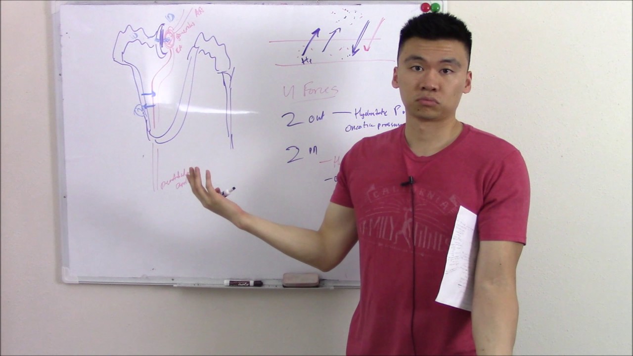
This video kicks off our renal block! While we will eventually talk all about the pathology, physiology, and pharmacology of the kidneys, we have to first start with some basic anatomy. So let's look at the kidneys. The outside portion of the kidney is called the cortex, while the inner portion is the medulla. There are small tubes that form the renal pyramid, which at the bottom is the renal papilla. These tubes drain into the small openings called minor calyces (calyx). These connect to larger openings called major calyces and eventually the renal pelvis and ureters. The ureters ultimately drain out into the bladder. Now know that your ureters go under the uterine arteries in women, vas deferens in men, and the iliac arteries in both. So there are three sites where urine flow can be obstructed. These sites are the ureteropelvic junction, pelvic inlet, and uretovesicular junction.
Now what is the blood supply of the kidneys? Arterial supply is via the renal artery and venous drainage is through the renal vein. Also important - your left gonadal vein joins the left renal vein. That does it for general anatomy for today! Now, let's talk about some physiology. We'll talk about urination.
The muscles of your bladder is called the detrusor muscle. Nothing leaks out because we have two sphincters, the internal sphincter and external sphincter, as well as the pelvic floor muscles that hold the urine back. Eventually, the stressed detrusor muscle sends signals to your brain that you need to urinate. Now your internal sphincter muscle is made of smooth muscle (involuntary), while your external sphincter is skeletal (voluntary). If the time is right, your external sphincter will open and you'll pee. Now if you urinate unintentionally, we call this urinary incontinence. The different types of urinary incontinence includes:
Stress: due to weak sphincter muscles or urethral hypermobility. Therefore, in stress like in coughing or straining, urine leaks. Treatment is kegel exercises, weight loss.
Urgency: due to overactive detrusor instability/hyperactivity. Treatment again is Kegels and antimuscarinics.
Overflow: incomplete voiding due to an outlet obstruction or detrustor underactivity. Often due to benign prostate hyperplasia, tumor and cancers, or neuropathy. That does it for this video! Next video we will talk about embryology!
Повторяем попытку...
Доступные форматы для скачивания:
Скачать видео
-
Информация по загрузке: