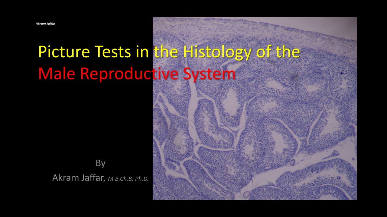
Picture tests in histology reproductive system - male
Автор: Human Anatomy Education
Загружено: 2016-05-10
Просмотров: 43964
Описание:
After completion of this session it is expected that the students will be able to identify, locate and describe the histological features of:
Male genital organs:
Seminiferous tubules: Spermatogenic cells: various stages of spermatogenesis and spermiogenesis: spermatogonia, spermatocytes and spermatogonia; Non-spermatogenic cells: Sertoli cells; Myofibroblasts and fibroblasts in the supporting tissue; Outline the structural changes in spermiogenesis; Sertoli cells: Describe their shape and position; Discuss their role in the formation of the blood-testis barrier; Describe the shape of the spermatozoon and its parts: head, neck and tail; Locate Leydig cells and outline their function.
Epididymis
Outline its function and relate structure to function: role of muscular layer and stereocilia; Describe the layers in its wall: epithelium and muscular layer.
Ductus (vas) deferens
Outline its function and relate structure to function: role of muscular layer and stereocilia; Describe the layers in its wall: epithelium and muscular layer.
Seminal vesicles
Describe the layers in its wall: epithelium and muscular layer and relate that to function; Identify the honeycombed appearance of its lumen and the foamy cells of mucosa; Give reason why it may contain spermatozoa.
Some images were cited in histology guide a virtual histology laboratory http://www.histologyguide.org/
Presented and edited by Akram Jaffar, PhD.
This video and its channel are supported by "Human Anatomy Education" Page on Facebook / anatomyeducation
Повторяем попытку...
Доступные форматы для скачивания:
Скачать видео
-
Информация по загрузке:


![Гистология яичников и овариальных фолликулов [Женская репродуктивная гистология. Часть 1 из 2]](https://imager.clipsaver.ru/cDs6goPOD1I/max.jpg)