How to do ct shoulder التصوير المقطعى للكتف
Автор: AHMED M SABER
Загружено: 2022-08-21
Просмотров: 3412
Описание:
#ct
#learning
#radiology
#siemens
#shortsvideo
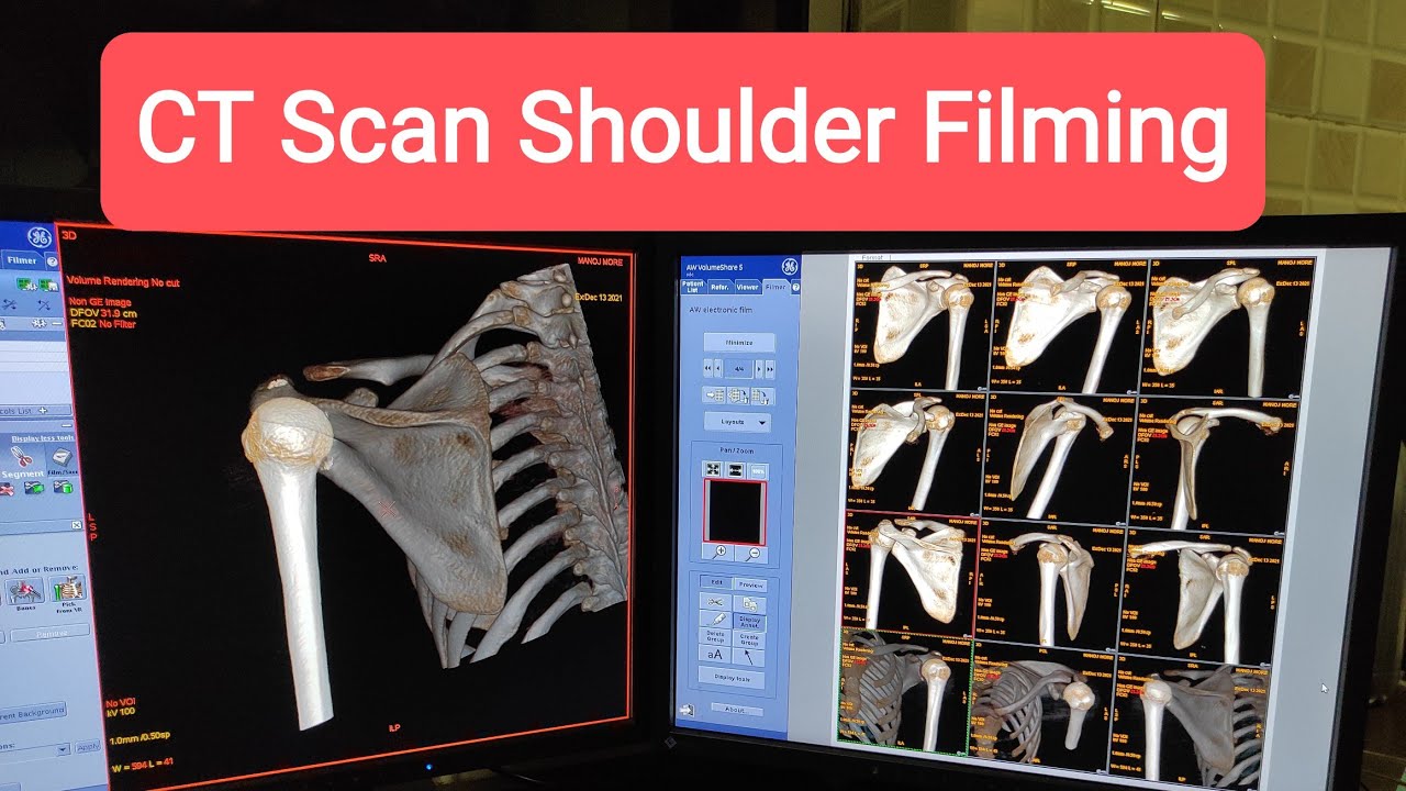
The CT shoulder protocol serves as an examination for the assessment of the shoulder joint. It is often performed as a non-contrast study. It can be combined with a CT arthrogram for the evaluation of labral injuries or the rotator cuff if MRI is contraindicated or in a postoperative setting where metallic implants are present.
Note: This article aims to frame a general concept of a CT protocol for the assessment of the shoulder. Protocol specifics will vary depending on CT scanner type, specific hardware and software, radiologist and perhaps referrer preference, patient factors e.g. implants, specific indications.
A typical CT of the shoulder might look like as follows:
Indications
Typical indications include the following 1-5:
proximal humeral fractures
scapular fractures
preoperative planning, e.g. for shoulder arthroplasty, Latarjet, remplissage
shoulder implants and complications
inflammatory or septic arthritis
bone and soft tissue tumors
image guidance
CT arthrography
if MRI is contraindicated or in the presence of metallic implants
glenoid labral tears or rotator cuff tears
Повторяем попытку...
Доступные форматы для скачивания:
Скачать видео
-
Информация по загрузке: