Tuberculosis (Tb), Tuberculosis x-ray, Tb chest x-ray.
Автор: Learn Radiology with Dr. Ben
Загружено: 2021-11-08
Просмотров: 1826
Описание:
✅ Click Now and Learn: Tuberculosis (Tb), Tuberculosis x-ray, Tb chest x-ray. Tuberculosis Radiology Findings (Chest-Spine-Peritoneum).
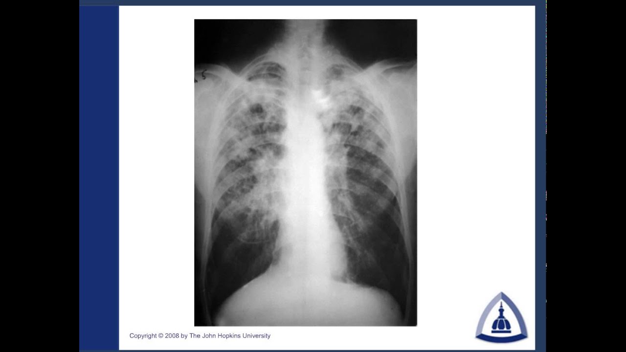
Primary pulmonary tuberculosis
In primary pulmonary tuberculosis, the initial focus of infection can be located anywhere within the lung and
has non-specific appearances ranging from too small to be detectable, to patchy areas of consolidation or
even lobar consolidation.
Radiographic evidence of parenchymal infection is seen in 70% of children and 90% of adults.
Tuberculosis (Tb), Tuberculosis x-ray, Tb chest x-ray.
00:01 Tuberculosis of the chest
06:05 Pott's Disease
09:07 Intraperitoneal Tuberculosis.
Cavitation is uncommon in primary TB, seen only in 10-30% of cases. In most cases, the infection becomes
localized and a caseating granuloma forms (tuberculoma) which usually eventually calcifies and is then
known as a Ghon lesion.
The more striking finding, especially in children, is that of ipsilateral hilar and contiguous mediastinal (paratracheal)
lymphadenopathy, usually right-sided.
This pattern is seen in over 90% of cases of childhood primary TB, but only 10-30% of adults.
These nodes typically
have low-density centers with rim enhancement on CT.
Occasionally these nodes may be large enough to compress adjacent airways resulting in distal atelectasis.
Tuberculosis (Tb), Tuberculosis x-rays, Tb chest x-ray.
Повторяем попытку...

Доступные форматы для скачивания:
Скачать видео
-
Информация по загрузке: