3D Reconstruction of ECoG Implanted Electrodes For Epilepsy Monitoring
Автор: Zack G
Загружено: 2015-03-14
Просмотров: 1304
Описание:
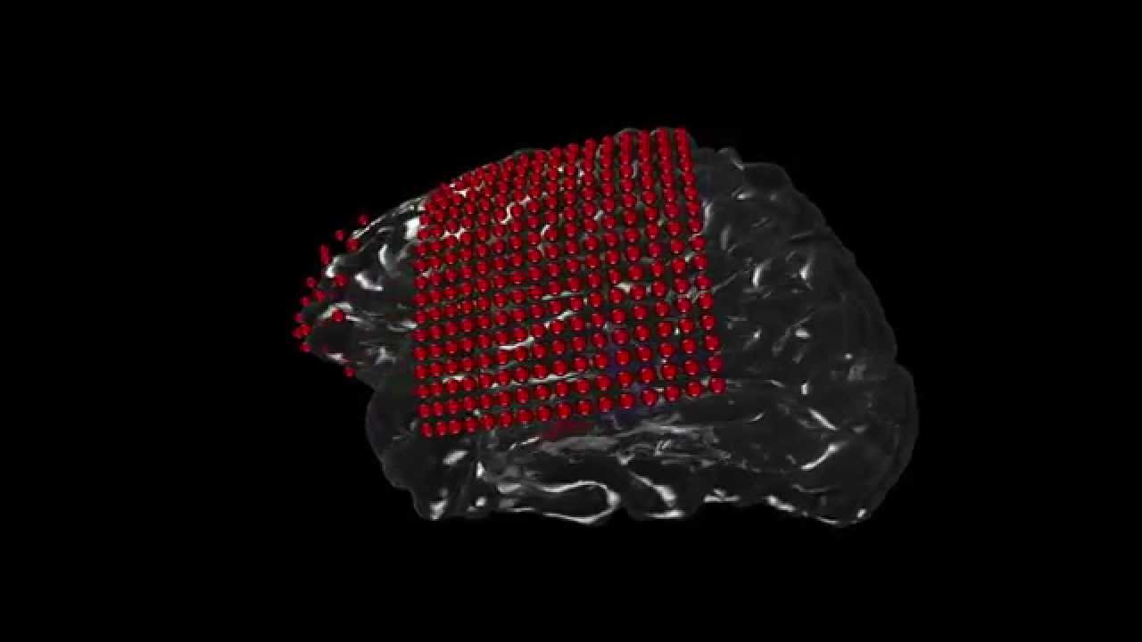
This 3D Cortical and Sub-cortical model was reconstructed from a patient's T1 Weighted MRI using freesurfer software (http://www.freesurfer.net/). The patient was implanted with intracranial (ECoG) electrodes for Epilepsy monitoring at UCSF hospital, and the electrode positions (red) were reconstructed using a high-resolution CT scan corregistered to the MRI.
The subcortical structures shown in the patient's right hemisphere are the hippocampus (blue), amygdala (yellow), globus pallidus (purple), putamen, and thalamus.
The code for visualization was written in C# for Unity 3D. Thanks to Roger Anguera for his help prototyping.
Повторяем попытку...
Доступные форматы для скачивания:
Скачать видео
-
Информация по загрузке: