Rotator Cuff tear Imaging
Автор: nabil ebraheim
Загружено: 2025-01-08
Просмотров: 8925
Описание:
Join this channel to support the clinic
/ @nabilebraheim
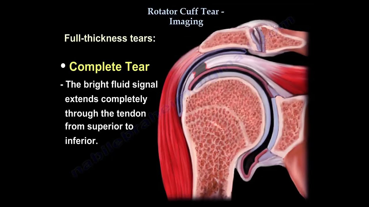
AP view x-ray: True AP (45° lateral) Patient can be standing, sitting, or lying down. Get a true AP view of the shoulder which will show the acromiohumeral interval which the normal interval should be between 7-14 mm. The x-ray may show proximal migration of the humeral head which means there is a chronic rotator cuff tea. Calcific tendonitis may also be seen on AP view x-ray. Beam aimed with 10° caudal tilt. Allows calcification of the acromion. The supraspinatus outlet view will show the shape of the acromion. OS acromial may be seen on axillary view x-ray. MRI is the best study for diagnosing rotator cuff tears. The MRI takes slices of cuts of the bone and the soft tissue structures as if you are taking a slice from an apple. The slices of the MRI can be seen in either a coronal, sagittal, or axial orientation. The normal rotator cuff appears dark on T1 and T2 on the MRI. Arthrogram is not used routinely. It is used when the patient is unable to have an MRI (pacemaker). Similar to a patient with a pacemaker that cannot have an MRI done of the spine (myelogram is done). Arthrogram will show the dye in the subacromial space. If the “Geyser Signs” is present with the dye leaking into the subacromial space as well as the AC joint, then there is a huge tear. If you add MRI to the arthrogram, that will increase the sensitivity and the specificity, and will show also any instability and partial tears better than arthrogram alone. When it comes to the MRI: the T2 is probably the best in showing the tears. The problem with MRI is the presence of asymptomatic cuff tear that are seen frequently in patients, especially older patients. 50% of asymptomatic patients 60 years or older will show rotator cuff tears on MRI. These tears increase in frequency with age. Rotator cuff tears and patient age is important because of a few points. 1- A lot of asymptomatic tears. 2- Patient is above the age of 40 years old with a dislocated shoulder may also have a rotator cuff tear. 3- Chronic tears are found in older patients which could involve multiple tendons. 4- The surgical outcome of rotator cuff repair depends on the age of the patient, the older the age of the patient, the worse the outcome. 5- Older patients with rotator cuff arthropathy can have reverse shoulder arthroplasty, which is different from younger patients. When you look at the MRI, you want to know the type of tear (partial or complete), the size of the tear (small, medium, or large). Rotator cuff tears are classified into either partial thickness or full thickness tears. - Partial thickness tears are classified by location into three types: • Articular –sided. • Bursal sided. • Intrasubstance. These tears are also classified according to the size of the tear (greater or less than 50% thickness). The fluid or dye will extend partially through the thickness of the tendon; however there is no retraction of the tendon. Full thickness tears: - Complete tear: the bright fluid signal extends completely through the tendon from superior to inferior. You want to know how much retraction is involved with the rotator cuff tear. Massive rotator cuff tears that are greater than 5 cm usually involve multiple tendons. The tendon will be retracted to the level of the glenoid and there will be atrophy of the muscle with fatty infiltration and probably superior migration of the humeral head. What is the quality of the muscle? Check for the presence of fatty atrophy on the sagittal view MRI. Normally the supraspinatus muscle occupies the fossa in the sagittal view. When the muscle is atrophied or abnormal, it does not occupy the fossa completely. Fatty muscle atrophy see on sagittal view usually means that there is either a chronic rotator cuff tear or suprascapular nerve injury. 0- Normal 1- Some fatty streaks 2- More muscle than fat 3- Equal fat and muscle 4- More fat than muscle It is important to know the proper orientation of the rotator cuff muscles in the sagittal view. Look at the long head of the biceps in an axial view. Check for subluxation of the biceps tendon which should mean there is also a tear of the subscapularis tendon. Remember: the long head of the biceps is anteriorly located. Look for cysts of the greater tuberosity on MRI which are seen in a lot of patients with rotator cuff tears. CT Arthtogram: - Done sometimes post operatively. - When the patient has anchors, we cannot see them well. - Some believe that ultrasound is better in these cases. - Used with frequency and is not expensive. - It may create dynamic evaluation. - There is a learning curve for using ultrasound imaging and it cannot evaluate the shoulder joint itself. - About 20% will show a rotator cuff tear in asymptomatic patients.
What percentage of asymptomatic patients above 60 years show rotator cuff tears on MRI?
10%
25%
50%
75%
Correct Answer: 50%
Повторяем попытку...
Доступные форматы для скачивания:
Скачать видео
-
Информация по загрузке:


