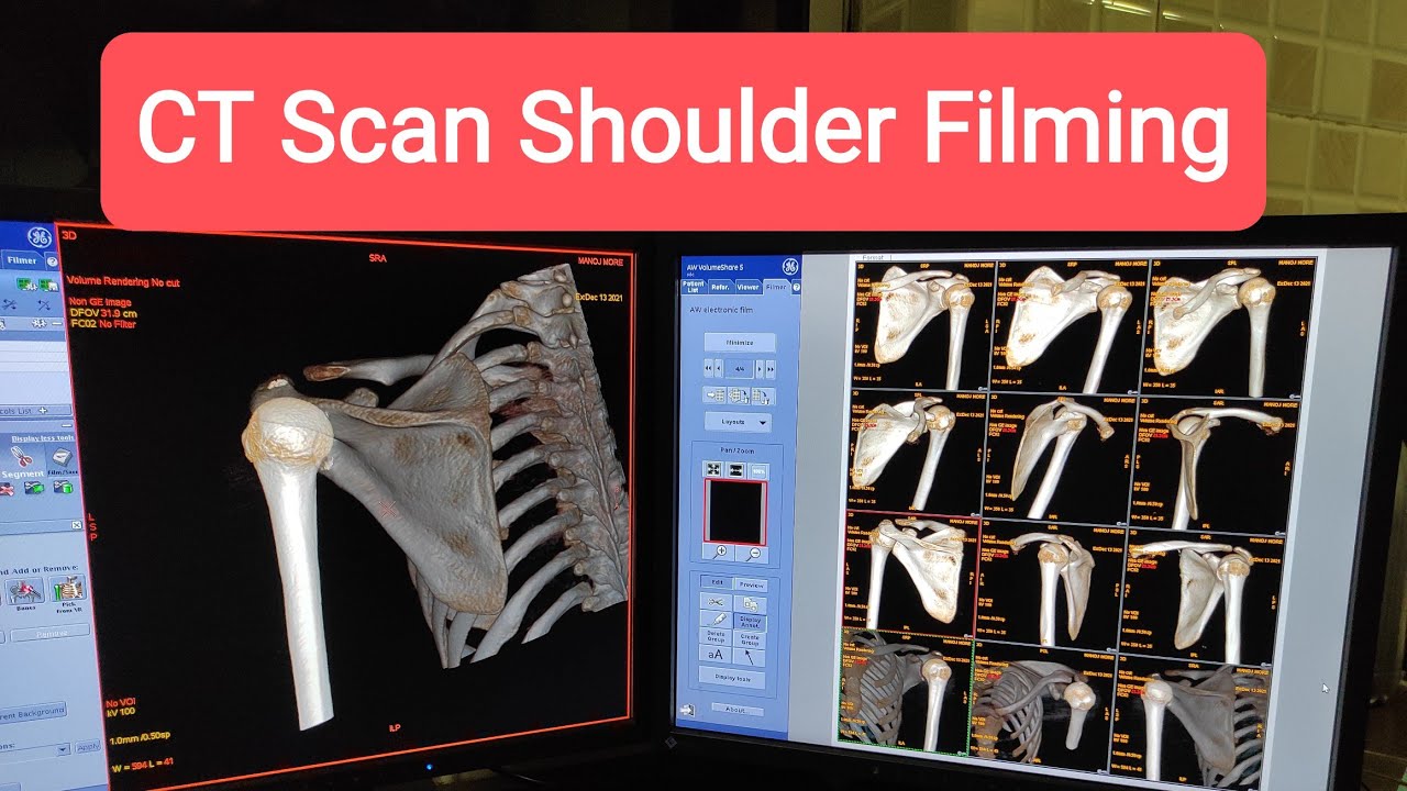
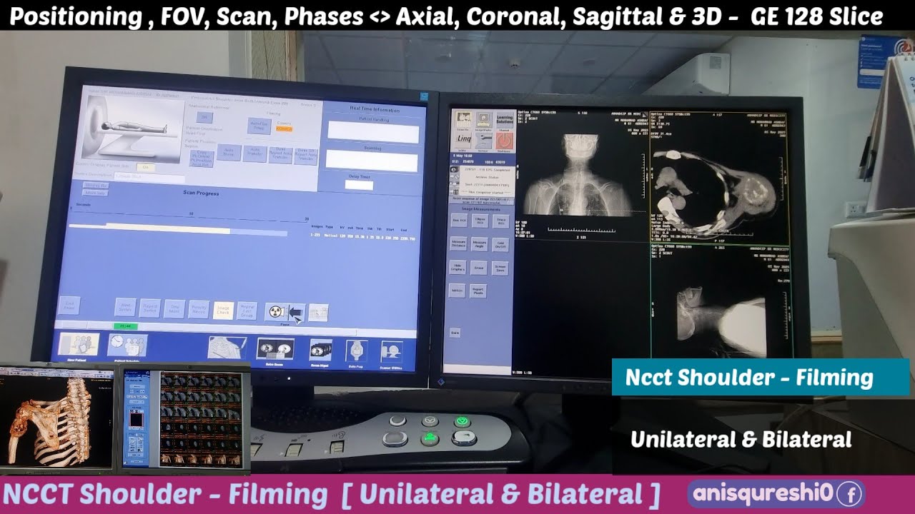
NCCT Shoulder | Positioning, FOV, Procedure & Filming | By Anis Qureshi
Автор: Anis Qureshi
Загружено: 2025-05-13
Просмотров: 1195
Описание:
In this video, explains the Non-Contrast CT (NCCT) Shoulder procedure in detail — from correct patient positioning to optimal filming for diagnosis.
📌 Step-by-Step Procedure:
1️⃣ Patient Preparation:
Remove metallic objects (necklaces, clothing with zippers/buttons) from scan area.
Explain the procedure and stress the need to remain still.
2️⃣ Positioning:
Head-first supine on CT table.
Arm of interest positioned by the side (neutral) or slightly externally rotated if tolerated.
Shoulder centered in gantry isocenter.
Immobilization pads can be used to minimize motion.
3️⃣ FOV (Field of View):
Cover from above the acromioclavicular joint down to the proximal third of the humerus.
Ensure inclusion of scapula, clavicle (lateral), and glenoid.
4️⃣ Scan Parameters:
kVp: 120 (typical)
mAs: Adjust as per patient size
Slice thickness: 0.5–1 mm for high-resolution detail
5️⃣ Scanning:
Perform NCCT scan without contrast.
Thin slices for multiplanar reconstruction (MPR) and 3D volume rendering.
🎥 Filming & Reconstructions:
Axial Images: Assess glenoid, humeral head, and scapular fractures.
Coronal MPR: Evaluate rotator cuff area, glenoid fossa, and joint space.
Sagittal MPR: Useful for labrum assessment and bone continuity.
3D Volume Rendering: For fracture mapping, surgical planning, and patient education.
Bone Window: For fracture lines, bony lesions.
Soft Tissue Window: For muscles, tendons, and soft tissue swelling.
🔍 Diagnoses Possible with NCCT Shoulder:
Fractures of humeral head, glenoid, clavicle, scapula
Shoulder dislocation
Degenerative changes
Bone tumors/lesions
Post-operative implant evaluation
🎯 Who Should Watch:
Perfect for Radiology students, X-ray/CT Technicians, Orthopedic imaging trainees, JKSSB aspirants, and practicing Radiographers.
📌 Follow for more radiology tutorials:
Instagram: @anisqureshi0
🔖 Hashtags:
#NCCT #CTScan #NCCTShoulder #ShoulderCT #Radiology #Radiographer #CTPositioning #FieldOfView #BoneWindow #SoftTissueWindow #CT3D #RadiologyTutorial #OrthopedicCT #RadiologyStudents #AnisQureshi #JKSSB #CTTrauma #ShoulderFracture #RadiographerTraining #CTFilming #CTBoneWindow #CTSoftTissueWindow #CT3DReconstruction #CTProcedure
.
Pdf Notes and books 📚 - https://t.me/anisqureshi0
This is the detailed video regarding NCCT ELBOW Filming Axial, Coronal, Sagittal & 3D Functions .
I will make videos in sections as will .
All Books in pdf format 👇👇🧠
https://t.me/anisqureshi0
All Radiology Related Videos 👇👇👇🧠🧠
1. Basic Human Anatomy and Physiology| Animated videos |
• Basic Human Anatomy and Physiology
Positioning of Radiograph..
• Positioning of X-ray Radiograph
Basic Radiology Physics.
• Basic Radiological Physics
CT Scan Anatomy and Pathology.
• CT Anatomy And Pathologies.
Anatomy of X-ray/ Radiographs
• X-ray Anatomy of Radiographs.
MRI Basics.
• MRI Basics
Ultrasound Basics.
• Ultrasound
CT Scan procedures,👇👇👇
• CT scan Procedures
#radiology radiology #xray #radiologia #medicine #radiologist #medical #mri #radtech #xraytech #radiographer #doctor #healthcare #radiologytech #imaging #xrays #radiologylife #radiologystudent #ct #ctscan #medicalimaging #ultrasound #hospital #surgery #nurse #medstudent #xraystudent #covid #medicina #medicalstudent #pathology
Повторяем попытку...
Доступные форматы для скачивания:
Скачать видео
-
Информация по загрузке: