Do you control for phototoxicity?
Автор: Nanolive, Looking inside life
Загружено: 2019-01-09
Просмотров: 1066
Описание:
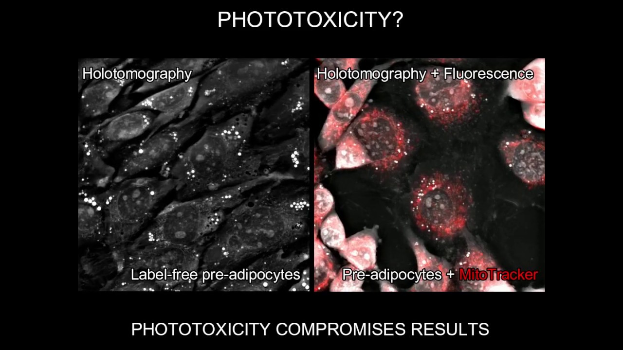
This video shows mouse Pre-adipocytes unlabeled and labeled with MitoTracker (dilution 1:5000) and have been imaged for 3 hrs at 1 image every 6s with holotomography (left panel) and holotomography + Cy5 fluorescence channel (right panel). We chose to use this type of cells because pre-adipocytes are known to be resistant and quite proliferative. Moreover, we use a mild fluorescence acquisition protocol, Cy5 is one of the least phototoxic light in fluorescence microscopy (long wavelenght). After less than an hour, cells on the right, exposed to fluorescence start dying through apoptosis. While, the ones on the left, imaged just through holotomography, show no sign of stress. This lack of phototoxicity can be even better appreciated in longer movies showing very sensitive cells like stem cells imaged for more than 50 hours as shown at this link: https://nanolive.ch/stem-cell-research/.
Technology: 3D Cell Explorer and 3D Cell Explorer-fluo microscope from Nanolive, Switzerland.
The 3D Cell Explorer is a high speed, high resolution and non-invasive live cell imaging microscope that can look deep inside biological systems. This allows you to record stunning 3D images of entire cells in just seconds and with a higher resolution than any conventional microscope on the market.
For this video, we used both types of microscopes (3D Cell Explorer and 3D Cell Explorer-fluo) to show the effects of phototoxicity.
For more information, visit nanolive.ch
Повторяем попытку...
Доступные форматы для скачивания:
Скачать видео
-
Информация по загрузке: