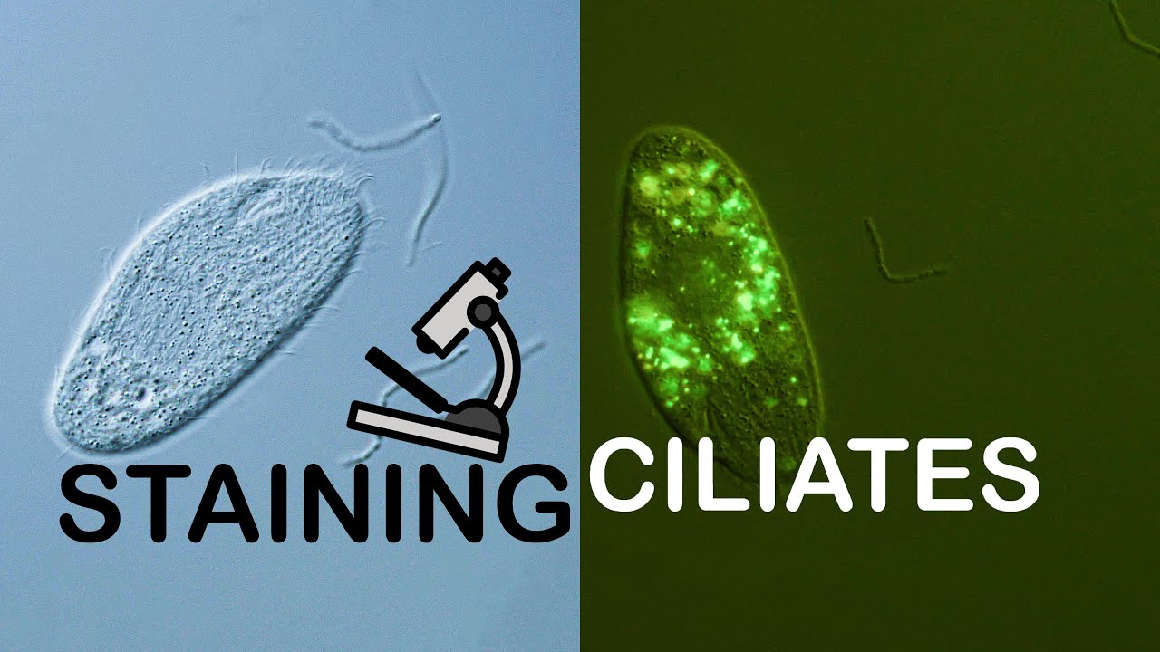
Staining Ciliates for fluorescent microscopy
Автор: VANOXmicroscopy
Загружено: 2021-03-30
Просмотров: 2122
Описание:
In this video I show the procedure of staining ciliates with acridin orange (AO) for fluorescent microscopy.
Acridin orange (AO) is a common fluorescent dye that stains DNA and also accumulates in acidic compartments. It's cell permeable and therefore allows live-cell imaging of the ciliates. The wavelength of the emitted light is dependent on the pH. It is therefore possible to observe the state of lysosomes and food vacuoles inside of the living organism as these compartments change pH during their life cycle.
The phototoxicity of AO is quite strong. The dye concentration and the intensity of the excitation light have to be balanced well for prolonged observation.
In this video I followed the AO dilution and staining procedure of Thilo Bauer which he describes in the following publication:
Thilo Bauer (2015), Interpretation der Acridinorange-Färbung von Ciliaten (Ciliophora) des Süßwassers und des Bodens
https://www.researchgate.net/publicat...
Dilution and staining protocol:
Dissolve 10mg of AO in 50ml of water
Make a second dilution by taking 10µl of the first dilution and pipeting it into 1ml of water. This is the final AO working dilution
Take 25µl of pond sample and mix it with 5µl of the final AO dilution
I used an Olympus Vanox AHBT3 with the original blue fluorescent cube together with a Cree XM-L2 LED as the excitation light source. I used the following objectives: Olympus SPlanApo 20/0,7, SPlanApo 60/1,4, UVFL 40/1,3 and a Leitz NPL Fluotar 50/1,0. The camera was a Panasonic S1 that is adapted using an Olympus NFK 2,5x photo eyepiece.
Background music:
Find Your Way Beat - Nana Kwabena
Into It - Kwon
Повторяем попытку...
Доступные форматы для скачивания:
Скачать видео
-
Информация по загрузке: