@Toppers Choice
Автор: @Toppers' Choice
Загружено: 2022-09-05
Просмотров: 157
Описание:
@Toppers Choice
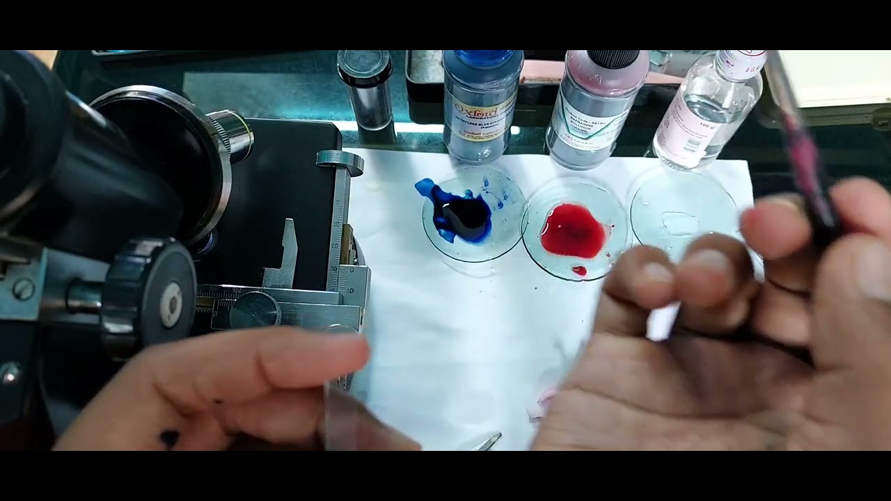
#ACTIVITY 9.5.2 (a) How to study plant cells?
What is required?
Onion bulb, slides, covers slips, forceps, needle, brush, watch
glass, glycerine, safranin/methylene blue and compound
microscope
How to proceed?
1. Take out a peel from the inner side of the fleshy scale leaf of
onion bulb.
2. Put the peel in watch glass containing water and cut it into
small rectangular pieces.
3. Transfer the cut pieces into another watch glass having
safranin/ methylene blue, and leave the peel for about 3
minutes.
4. Wash the peel with water to remove excess stain.
5. Put a drop of glycerine in the middle of a clean slide and
transfer the stained peel onto it.
6. Place the cover slip on the material by lowering it slowly with
the help of a needle.
7. Remove excess glycerine with blotting paper.
8. Keep the prepared slide on the stage of the microscope and
observe under low power.
9. Draw what you have observed under microscope and label
the diagram.
Fig. 9.5.2(a) Cells observed in an onion peel
What have you learnt?
1. The onion peel cells are rectangular in shape and compactly
arranged.
2. Each cell is surrounded by a thick cell wall.
3. A dense spherical body, the nucleus, is seen in each cell.
4. Cell membrane can also be observed
Precaution
While putting the cover slip care should be taken to avoid the
entry of air bubbles. For this purpose keep the cover slip at 45
degree on the slide and then slowly lower it using the needle.
Extension
Observe leaf epidermal peel of Tradescandia/Rhoeo/
Bryophyllum, onion root tip.
Повторяем попытку...
Доступные форматы для скачивания:
Скачать видео
-
Информация по загрузке: