Histology of Tooth Development : Shotgun Histology
Автор: Dr.G.Bhanu Prakash
Загружено: 2019-05-14
Просмотров: 9871
Описание:
📌 𝐅𝐨𝐥𝐥𝐨𝐰 𝐨𝐧 𝐈𝐧𝐬𝐭𝐚𝐠𝐫𝐚𝐦:- / drgbhanuprakash
📌𝗝𝗼𝗶𝗻 𝗢𝘂𝗿 𝗧𝗲𝗹𝗲𝗴𝗿𝗮𝗺 𝗖𝗵𝗮𝗻𝗻𝗲𝗹 𝗛𝗲𝗿𝗲:- https://t.me/bhanuprakashdr
📌𝗦𝘂𝗯𝘀𝗰𝗿𝗶𝗯𝗲 𝗧𝗼 𝗠𝘆 𝗠𝗮𝗶𝗹𝗶𝗻𝗴 𝗟𝗶𝘀𝘁:- https://linktr.ee/DrGBhanuprakash
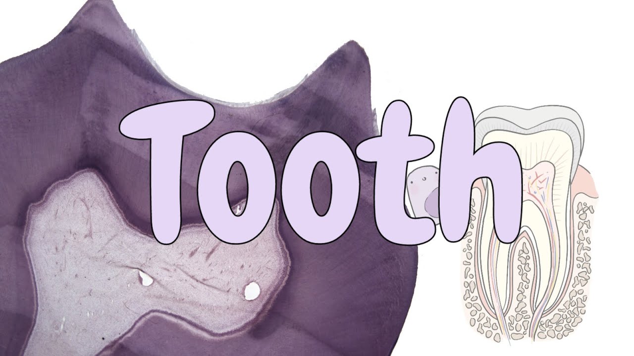
Histology of Tooth Development: Shotgun Histology
Tooth development is the complex process by which teeth form from embryonic cells, grow, and erupt into the mouth. Although many diverse species have teeth, non-human tooth development is largely the same as in humans. For human teeth to have a healthy oral environment, enamel, dentin, cementum, and the periodontium must all develop during appropriate stages of fetal development. Primary (baby) teeth start to form between the sixth and eighth weeks in utero, and permanent teeth begin to form in the twentieth week in utero. If teeth do not start to develop at or near these times, they will not develop at all.
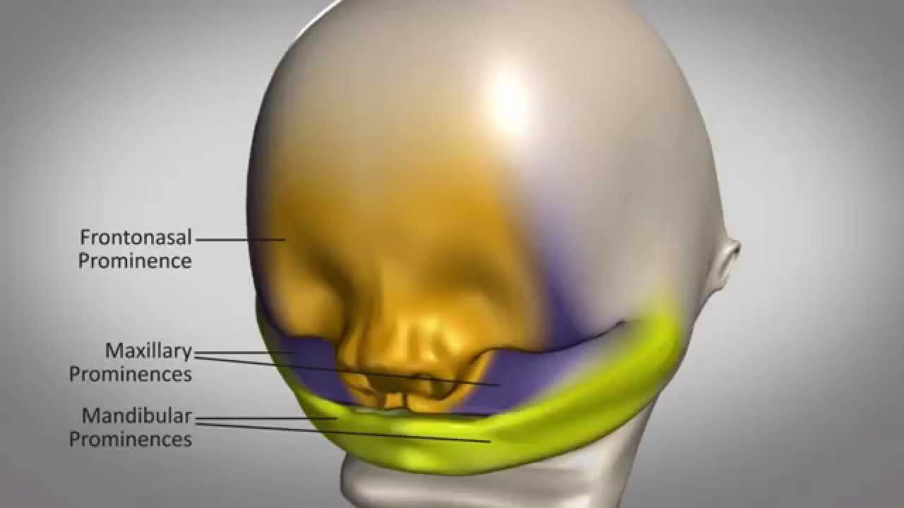
A significant amount of research has focused on determining the processes that initiate tooth development. It is widely accepted that there is a factor within the tissues of the first branchial arch that is necessary for the development of teeth
The tooth bud (sometimes called the tooth germ) is an aggregation of cells that eventually forms a tooth. These cells are derived from the ectoderm of the first branchial arch and the ectomesenchyme of the neural crest. The tooth bud is organized into three parts: the enamel organ, the dental papilla and the dental follicle.
The enamel organ is composed of the outer enamel epithelium, inner enamel epithelium, stellate reticulum and stratum intermedium. These cells give rise to ameloblasts, which produce enamel and the reduced enamel epithelium. The location where the outer enamel epithelium and inner enamel epithelium join is called the cervical loop. The growth of cervical loop cells into the deeper tissues forms Hertwig's Epithelial Root Sheath, which determines the root shape of the tooth.
The dental papilla contains cells that develop into odontoblasts, which are dentin-forming cells. Additionally, the junction between the dental papilla and inner enamel epithelium determines the crown shape of a tooth. Mesenchymal cells within the dental papilla are responsible for formation of tooth pulp.
The dental follicle gives rise to three important entities: cementoblasts, osteoblasts, and fibroblasts. Cementoblasts form the cementum of a tooth. Osteoblasts give rise to the alveolar bone around the roots of teeth. Fibroblasts develop the periodontal ligaments which connect teeth to the alveolar bone through cementum.
#histologyoftoothdevelopment #toothdevelopmenthistology #toothdevelopment #histologytoothdevelopment #developmentoftoothhistology
Повторяем попытку...

Доступные форматы для скачивания:
Скачать видео
-
Информация по загрузке:


![Skin Histology [Integumentary System Histology Part 1 of 2]](https://imager.clipsaver.ru/NBEjKRuGHjY/max.jpg)


![Хирургическая анатомия шейной диссекции: часто задаваемые вопросы и ответы [201] Дидактика](https://imager.clipsaver.ru/oSQLNfo4rXs/max.jpg)

![Гистология слюнных желез [Гистология ЖКТ 3 из 4]](https://imager.clipsaver.ru/larjdAJN9Js/max.jpg)
