Histology of spleen
Автор: Knowing Anatomy
Загружено: 2020-09-11
Просмотров: 114481
Описание:
The spleen is located in the left upper quadrant region of the abdomen. More precisely, it is found posterior to the stomach and anterior and inferior to the left hemidiaphragm at the level of ribs 9-10. On the medial side, the spleen is in relation to the left kidney and inferiorly it sits on the left colic flexure (splenic flexure).
By being almost an entirely intraperitoneal organ, the spleen is mobile within the abdominal cavity. The hilum of the spleen is the only part of the spleen that is peritoneum free.
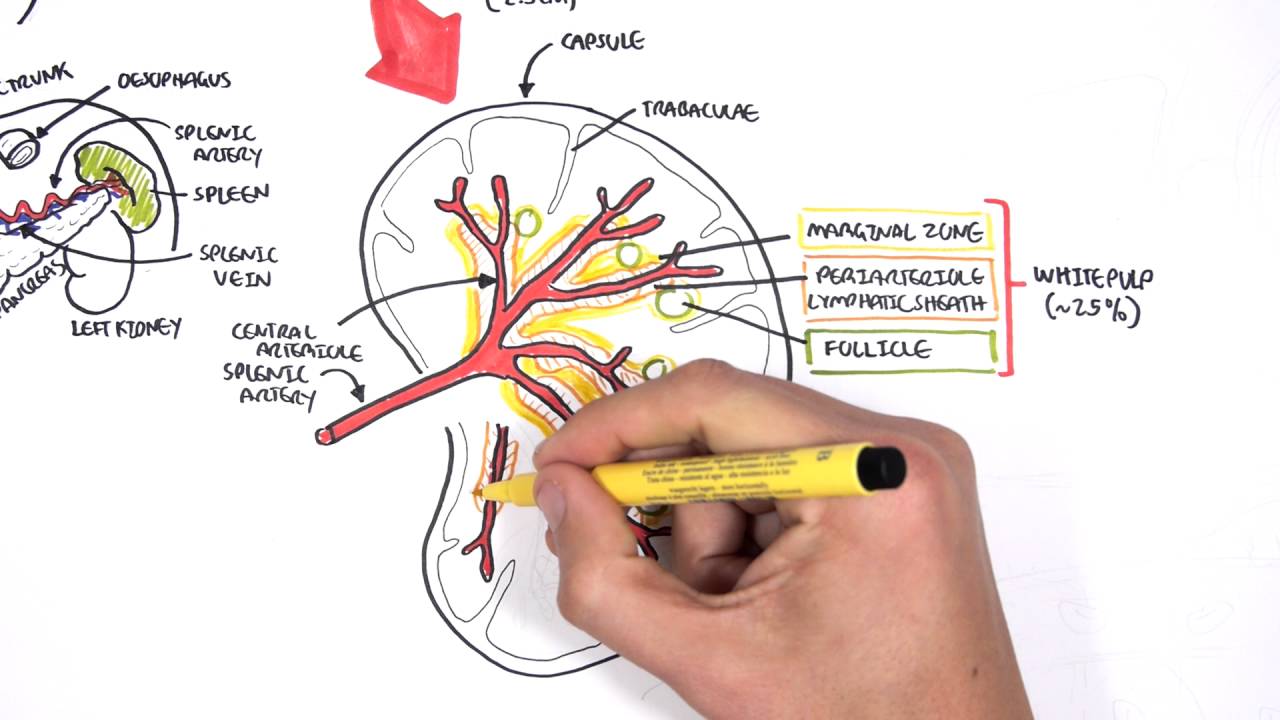
Structure
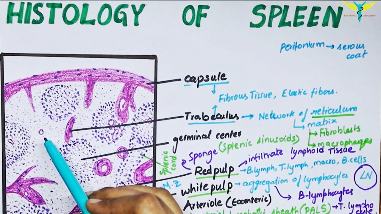
Being an intraperitoneal organ, the spleen is covered by a layer of visceral peritoneum. Underneath the peritoneum is the capsule of the spleen, encasing its parenchyma.
The capsule of the spleen consists of dense irregular fibroelastic tissue. The connective tissue of the capsule contains contractile cells called myofibroblasts. By producing weak contraction of the capsule, these cells help to discharge the blood stored within the spleen into the circulation. The capsule also allows the spleen to significantly increase in size when necessary and discharge a large amount of blood to contribute to the tissues oxygenation, like during physical exercise. At the level of the hilum, the capsule splits into several septae called trabeculae which penetrate into the parenchyma of the spleen and partly divide its tissue.
Normal spleen histology (diagram).
Like every other organ, the spleen consists of stroma and parenchyma. The stroma of the spleen is composed mainly of a network of reticular connective tissue. This mesh provides support for blood cells and cells of the immune system (lymphocytes, macrophages, and dendritic cells). The parenchyma of the spleen is divided into two functionally and morphologically distinct compartments (red pulp and white pulp) divided by a tissue layer called the marginal zone. Outside the marginal zone is the perifollicular zone which contains sheathed capillaries and blood-filled spaces without endothelial lining.
Red pulp
The red pulp occupies the majority of the stromal tissue of the spleen. It consists of the cords of Billroth and splenic sinusoids. The cords of Billroth (splenic cords) are the cellular aggregations supported by the reticular connective tissue. They appear as stripes and consist of of macrophages, plasmocytes and blood cells.
Splenic sinusoids are found between the cords of Billroth. They are filled with blood and give the red pulp its distinguishable red appearance. Blood slowly flows through the sinusoids where it is exposed to macrophages from the cords of Billroth, patiently waiting for foreign antigens that can appear in the blood. In a nutshell, the red pulp functions as a blood filter for various toxins, destroying them before they enter systemic circulation and get the chance to spread throughout the body and damage other organs.
White pulp
The white pulp of the spleen is made of three different compartments: Periarterial lymphatic sheath (PALS), lymphatic follicles and the marginal zone.
The PALS consists of a central artery (a branch of the splenic artery) surrounded by a sheath of lymphoid tissue. Here, the lymphoid tissue organized into two layers: The inner layer and outer layer. The inner layer is mainly composed of T lymphocytes which is why it is also called the T-zone. The outer layer has a more diverse cellular morphology, containing T and B lymphocytes.
The branches of central arterioles are surrounded by the sharply defined areas of B lymphocytes, comprising the lymphatic follicles of the spleen. There are two types of lymphatic follicles depending on the features of the B lymphocytes that comprise them: Primary follicles and secondary nodules.
The marginal zone is found on the very edge of the lymphatic follicles, containing different immune cells that are well equipped for starting an appropriate immune response.
Blood vessels
Spleen blood vessels and microcirculation (overview diagram)
Since the spleen is a blood filter, one has to assume that it is a highly vascular organ. Blood from the splenic artery enters the spleen through the hilum. From there, the artery divides into smaller branches that enter the splenic parenchyma following the course of trabeculae. Together with the trabeculae, the arteries branch throughout the parenchyma and gradually decrease in diameter. Eventually, smaller arterioles leave the dense connective tissue of trabeculae entering the parenchyma, where they are surrounded by PALS.
Повторяем попытку...
Доступные форматы для скачивания:
Скачать видео
-
Информация по загрузке: