3D visualization of hindbrain-spinal circuit in larval zebrafish
Автор: Discover Magazine
Загружено: 2013-03-18
Просмотров: 6049
Описание:
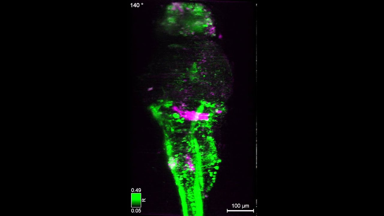
Scientists at the Howard Hughes Medical Institute studied live zebrafish larvae that had been genetically encoded with a calcium indicator called GCaMP5G. They suspended the larva in a gel and then beamed it with lasers. Just before a neuron fires, its action potential is expressed via a spike in calcium ions, so when one of the genetically modified larva's neurons reached its action potential, it glowed. This showed the researchers the firing of the neurons without them having to attach a bunch of electrodes to the fish.
Over the course of an hour the researchers used laser beams to scan the larva every 1.3 seconds, exciting the retina of the zebrafish with each scan. This microscopy method allowed the researchers to record up to 80 percent of the fish's 100,000 neurons at single-cell resolution.
This is the first time scientists have recorded such a high percentage of an organism's brain activity at such a high resolution.
Read the full story at: https://www.discovermagazine.com/plan...
—
Courtesy of Misha B. Ahrens and Philipp J. Keller, Howard Hughes Medical Institute.
—
ABOUT DISCOVER:
Discover is your source for science that matters, makes sense and is a lot of fun, too.
Visit us at: http://www.discovermagazine.com/
FOLLOW US:
Facebook: / discovermag
Twitter: / discovermag
Instagram: / discover.magazine
SUPPORT US:
Subscribe to Discover: https://bit.ly/34seAFT
Sign up for our newsletter: https://www.discovermagazine.com/news...
My Science Shop: https://www.myscienceshop.com/
STAY CURIOUS!
Повторяем попытку...
Доступные форматы для скачивания:
Скачать видео
-
Информация по загрузке:


