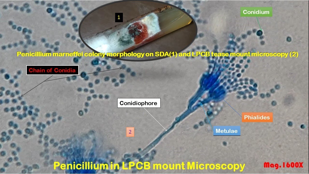
Penicillium marneffei colony morphology on SDA and LPCB tease mount Microscopy
Автор: MedLabSolutions
Загружено: 2022-01-27
Просмотров: 488
Описание:
Penicillium marneffei colony morphology on SDA,
Penicillium marneffei LPCB tease mount Microscopy,
conidia, phialides, septate hyphae, and conidiophore of Penicillium marneffei,
Penicillium marneffei colony morphology on SDA and LPCB tease mount Microscopy,
conidia of Penicillium,
phialides of Penicillium,
septate hyphae of Penicillium,
conidiophore of Penicillium,
Atlas of Fungi: Introduction, List of Contents, and Description @https://medicallabnotes.com/atlas-of-...
India Ink Preparation of CSF for observation of capsules of Cryptococcus neoformans in case of Cryptococcal meningitis
India Ink Preparation of CSF Positive
Cryptococcus capsules in Nigrosin preparation
Cryptococcus colony characteristics on Sabouraud dextrose agar
Cryptococcus neoformans mucoid colony on SDA
Cryptococcus Growth on Sabouraud Dextrose Agar (SDA) showing smooth, mucoid, and cream-colored colonies
Live and Dead Cryptococcus neoformans Footages in Methylene Blue wet mount
Live and dead Cryptococcus yeast cells under the Microscope
Yeast cells of Cryptococcus neoformans in Gram-stained smear of culture
India Ink Preparation from the culture of Cryptococcus neoformans
Cryptococcus capsules in Giemsa stained smear
Cryptococcus neformans in LPCB tease mount of culture
Cryptococcus in a saline wet mount of culture
Birdseed agar showing colony characteristics of Cryptococcus neoformans
Candida and Cryptococcus growth on Coffee agar
Cryptococcus neoformans mucoid strain growth on MacConkey medium from CSF
Cryptococcus neoformans mucoid strain growth on blood agar from CSF
Urease Positive Cryptococcus neoformans Demonstration
Candida albicans and Cryptococcus neoformans growth on SDA
Candida albicans colony characteristics on SDA
Candida colony morphology on Sabouraud dextrose agar (SDA)
Gram Positive Yeast cells of Candida
Yeast cells of Candida in saline wet mount of culture
Germ tube test positive Candida albicans
Chlamydospores of Candida albicans Demonstration
Antifungal Drugs Susceptibility Testing (AFST) pattern of Candida tropicalis
Candida tropicalis yeast cells in Gram stained smear of culture
Candida tropicalis yeast cells in saline wet mount of culture
KOH mount of ear discharge showing fungal elements-Septate hyphae with V-shaped or acute-angle or dichotomous branching
Numerous conidia of Aspergillus and pus cells in Gram-stained smear of CSOM patient ear discharge
Aspergillus niger colony morphology on Sabouraud Dextrose Agar (SDA)
Aspergillus niger colony characteristics on SDA
Conidia, phialides, vesicle, stipe and conidiophore of Aspergillus in LPCB tease mount microscopy
Aspergillus niger colony characteristics on Cornmeal Agar (CMA)
A beautiful Aspergillus niger colony characteristics on SDA demonstration
Aspergillus fumigatus fungal elements in KOH mount of aural discharge microscopic footage
Aspergillus fumigatus colony morphology on Sabouraud Dextrose Agar (SDA)
Aspergillus fumigatus mycelium, hyphae, and conidia in LPCB tease mount
Aspergillus fumigatus colony morphology on SDA
Fungal hyphae and conidia in KOH mount of Pus
Aspergillus flavus Colony Characteristics on Czapek Dox Agar
Aspergillus flavus Colony Characteristics on CMA
Conidia, phialides, vesicle, stipe, and conidiophore of Aspergillus flavus in LPCB tease mount microscopy
Bipolaris Colony Morphology on SDA
Bipolaris species in LPCB tease mount showing sympodial development of pale brown pigmented, pseudseptate conidia on a geniculate or zig-zag rachis
Cladosporium Colony Morphology on SDA
Cladosporium reverse pigment on SDA
Trichophyton mentagrophytes Colony characteristics on SDA
Trichophyton mentagrophytes Colony Morphology on SDA (plate) and SDA (tube)
LPCB tease mount ofTrichophyton mentagrophytes culture showing microconidia (1), macroconidia (2), hyphae (3), chlamydospores(4), and spiral hyphae(5)
Ringworm due to Trichophyton rubrum
Trichophyton rubrumcolony morphology on SDA
Trichophyton rubrum microconidia, macroconidia and hyphae in LPCB tease mount microscopy
Microsporum ferrugineum Colony Morphology on SDA
Microsporum ferrugineum in LPCB tease mount microscopy
Penicillium colony morphology on SDA
Penicillium cheresanum in LPCB tease mount exhibiting long single celled conidia
Penicillium marneffei colony morphology on SDA
Penicillium marneffei diffusible brownishred to wine-red pigment on SDA
Penicillium marneffei fungal elements in LPCB teast mount microscopy
Paecilomyces marquandii Colony Morphology on CMA
Conidiophores, phialides and conidia of Paecilomyes marquandii in LPCB Tease mount
Sporangiophores, columellae and rhizoids of Rhizopus in LPCB mount
Mucor causing Mucormycosis colony morphology
Sporangium, sporangiospores, columella, sporangiophore or aerial hyphae of Mucor species in LPCB tease mount
Повторяем попытку...
Доступные форматы для скачивания:
Скачать видео
-
Информация по загрузке:


![Dimorphic Fungi: Paracoccidioidomycosis and Penicilliosis [Hot Topic]](https://imager.clipsaver.ru/u-IR6LKsk-U/max.jpg)