Lecture On Microscopy Principles (Resolution, NA) & Types: Brightfield, Darkfield, TEM, SEM | MBBS
Автор: Santiniketan Medical College
Загружено: 2025-12-03
Просмотров: 78
Описание:
This comprehensive Microbiology lecture provides an in-depth exploration of the Microscope—from its basic principles and historical development to the working mechanics and classification of modern systems. Essential for MBBS and life science students, this class focuses particularly on the critical concepts of Magnification and Resolution and details the diverse applications of light and electron microscopy.
Key Topics and Time-stamped Chapters (Use these for quick navigation):
Time Topic
[00:00] Etymology of Microscope and the overall process of Microscopy.
[00:40] Brief History of Microscope Development (Hans & Janssen, Leeuwenhoek, Hooke).
[02:09] Classification of Modern Microscopy (Optical/Light, Electron, Scanning Probe).
[03:00] Parts of a Compound Light Microscope (Light Source, Condenser, Stage, Lenses).
[05:23] Key Parameters of Microscopy: Magnification vs. Resolution (D).
[06:17] Understanding Resolution: Relationship between Resolution (R) and Limit of Resolution (D).
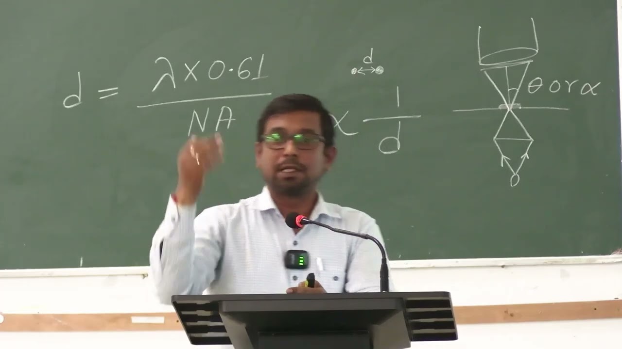
[08:23] The Resolution Formula: D=0.61×λ/(N×sinα).
[10:00] Numerical Aperture (NA) and the role of Immersion Oil (Cedar Root Oil) in increasing the Refractive Index (N) for better resolution.
[12:17] How to achieve Higher Resolution (Lower Wavelength, Higher NA).
[13:26] Principles of Magnification (Real, Inverted, Virtual Images).
[16:05] Types of Light Microscopy
[16:05] Brightfield Microscopy: Bright background, dark (stained) object.
[16:52] Dark Field Microscopy: Uses Annular Ring. Used for living organisms (e.g., flagella, cilia); bright object on dark background.
[19:51] Phase Contrast Microscopy: Uses a Phase Ring for even better contrast and resolution of transparent specimens.
[22:15] Fluorescence Microscopy: Uses UV Light and Fluorophores (dyes). Principles of electron excitation/emission and use of Dichroic Mirrors/Filters.
[26:47] Confocal Microscopy: Uses Laser light and a Pinhole Filter to eliminate out-of-focus light for sharp 3D imaging.
[28:18] Electron Microscopy (Uses Electrons, not Light)
[28:20] Transmission Electron Microscope (TEM): Used for internal structure (requires ultra-thin sections).
[28:28] Scanning Electron Microscope (SEM): Used for 3D external structure (surface morphology).
[30:35] Atomic Force Microscopy (AFM): Used to visualize atomic-level structures.
SANTINIKETAN MEDICAL COLLEGE & HOSPITAL
Real Classroom Demonstration/Lecture
On: MICROBIOLOGY by Dr. Chinmoy Bandopadhyay
(MBBS Batch: 2024-25)
Conducted on: 04-12-25
Повторяем попытку...
Доступные форматы для скачивания:
Скачать видео
-
Информация по загрузке: