TPLO Info Step 3: Proper Diagnosis
Автор: TPLO Info
Загружено: 2018-10-19
Просмотров: 3759
Описание:
To see the original post on Step 3: Proper Diagnosis visit - https://tploinfo.com/6-steps/step-3/
Transcription -
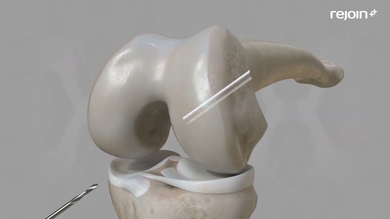
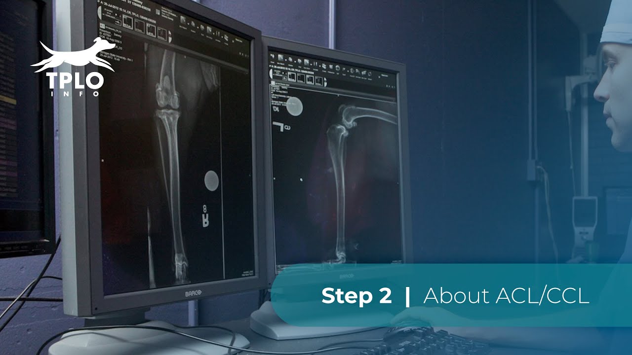
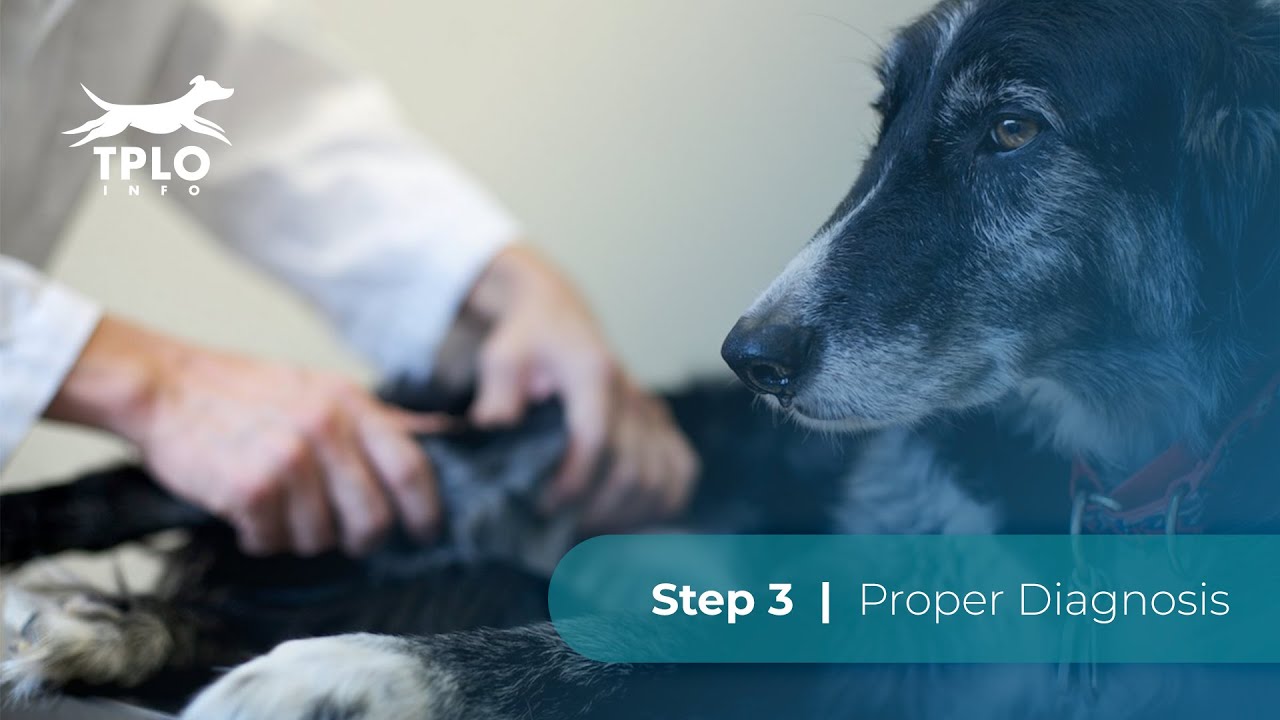
Okay step three, how do we diagnose an ACL injury in the dog? Well, there's several ways and the first thing we're gonna talk about is is, how do we diagnose it just on a direct physical examination? Followed by, how do we diagnose it using x-rays? And then lastly, what are some other signs or indicators that we maybe able to pick up on to figure out weather or not a dog has either an early partial ACL injury or a complete ACL injury. An important thing to consider before we play this video is that some reports are online that these tests may hurt your dog's knee. You'll se in this test, that this is no different than your pet taking a step. So, it only hurts your dog's knee, if at all as much as them taking a step. We're gonna play this video and as we do, watch the movement between the femur and the tibia. The hands in this video are placed, one on the femur and two on the tibia. And so, it's just like the x-rays you've been looking at from the side view. As this video plays, you can see that the two bones are moving with respect to one another. This is called the direct drawer test. The second video we have here, is called the indirect drawer test, or cranial-tibial thrust. This is the notion that's produced when the dog takes a step and weight travels through the leg. We alluded a little bit to this motion In step two, you can se force coming up through the tibia and producing instability in this dog's knee. This dog has a severe, complete ACL tear. Here's another video of the tibial compression test or indirect drawer test, using continuous x-ray. On continuous x-ray, the tibia is moving forward with respect to the femur as weight is transferred through the leg. We want to give you an idea of what x-rays look like. And how does your veterinary know, based on an x-ray that your dog may have an ACL tear. Now, the only real way to know, there's an ACL tear is to either feel the instability, see that the two bones have moved with respect to one another or look at it with your own eyes during surgery. In the x-ray on the left hand side, the knee is normal, there is no joint affusion and no arthritis, in the x-ray on the right, there's a light gray patch within the joint space that is joint affusion or swelling. There's also small bone spears or osteophytes on the patella which you know is another name for the knee cap. Here are three side x-rays of the dog's knee. The image on the left is normal, the image in the middle is moderate arthritis and swelling associated with either a partial or complete ACL tear. The image on the right is most likely associated with a complete ACL tear as the arthritis and swelling are now severe. Other indicators that a dog may have a partial or complete ACL tear include atrophy, with is a loss of muscle, you can notice that one leg is smaller in diameter than the other, effusion, which can be felt usually by a veterinarian, buttress which is a swelling on the inside of the dog's knee or medio buttress is another term for that, meaning that on the inside of the dog's knee, there's excessive scar tissue as the body's attempt to stabilize the unstable ACL injury. Finally some veterinarians can feel trochlear osteophytes, which are bone spears by common term, there can be pain during movement of your dog's knee on examination, especially when we extend the dog's knee and then finally some owners can literally hear an audible clicking noise, coming from their dog's knee. Most often, this is the result of a meniscal tear, we talked about in earlier videos that the meniscus can become injured once the ACL is torn.
Повторяем попытку...
Доступные форматы для скачивания:
Скачать видео
-
Информация по загрузке: