Vascular Imaging of the Head and Neck - Case D
Автор: LearnNeuroradiology
Загружено: 2021-08-10
Просмотров: 8336
Описание:
This case is the fourth and final case that goes with the vascular capstone course you can find here:
https://bit.ly/CTAcourse
On that page, there is a scrollable case that you can go through to teach you how to approach a CTA of the head and neck in a real patient.
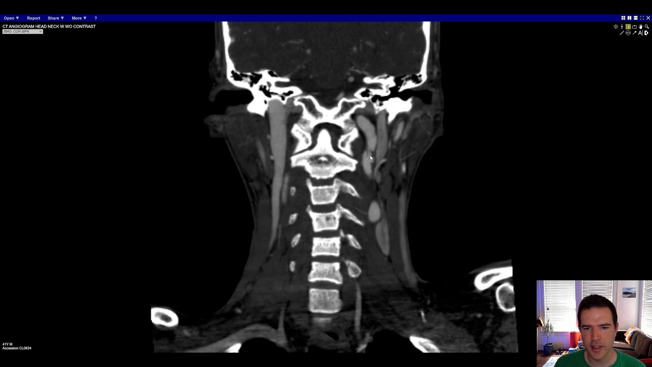
This case is a 41 year-old man after a trauma in a motor vehicle collision (MVC). Take a look and see what you think before continuing on ( https://bit.ly/CTAcaseD).
Starting with the CTA of the neck, this is not a normal case. If you follow your normal search pattern, you will see that there are a number of abnormalities, starting from the right internal carotid artery (ICA), which is lumpy and irregular looking. The left ICA is worse, with areas of narrowing, outpouchings, and linear filling defects that represent little areas of the intima that are lifted up by trauma. The little outpouchings along the margins of the vessel are little pseudoaneurysms, or areas where the vessel is injured and contrast is able to leak out into the surrounding area of damaged vessel. Both vertebral arteries are also abnormal with multifocal irregularity and a small pseudoaneurysm on the right.
This is a dramatic example of traumatic vascular injury in the neck. After high energy or penetrating (think gunshot or stab wound), the great vessels can be injured and jeopardize the blood supply to the brain. These injuries are graded on the Denver, or Biffl, scale which ranges from 1-5. You can read more about it here (https://radiopaedia.org/articles/biff.... Injuries to the vessels in the neck are most commonly pseudoaneurysms, meaning that one layer or more of the vessel is injured and the wall of the aneurysm does not contain all the layers (intima-media-adventitia). Contrast this to intracranial aneurysms, which are true aneurysms and the wall contains all the layers.
This is the last of the case examples for the vascular capstone course. If you haven't already, I recommend going back to the vascular capstone course, where you can review the other browseable cases with explanations. The capstone overview is at https://bit.ly/CTAcourse, if you'd like to see all the cases and videos.
Or, see all of the vascular capstone videos in the vascular imaging capstone playlist.
Check out this video and additional content on http://www.learnneuroradiology.com
Повторяем попытку...
Доступные форматы для скачивания:
Скачать видео
-
Информация по загрузке:


![[CT] Neck | Search Pattern](https://image.4k-video.ru/id-video/1_Z6FT4JjS0)


