human heart structure and function
Автор: Visible Science
Загружено: 2023-01-09
Просмотров: 309306
Описание:
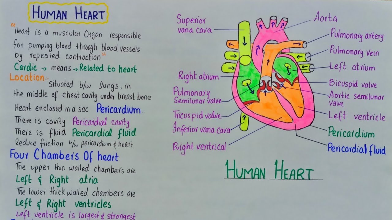
HUMAN HEART
Heart is a muscular organ responsible for pumping blood through blood vessels by repeated
contractions. The term “cardiac” means “related to the heart”. The bulk of the walls of heart chambers
is made of cardiac muscles.
In human body, heart is situated between lungs, in the middle of chest cavity (thorax) under
breastbone.
Heart is enclosed in a sac known as pericardium. There is a fluid, known as pericardial fluid,
between pericardium and heart walls. It reduces friction between pericardium and heart, during
heart contractions.
Human heart consists of four chambers, like the heart of birds and other mammals. The upper
thin-walled chambers are called left and right atria (singular ‘atrium’), and the lower thick-walled
chambers are called left and right ventricles. Left ventricle is the largest and strongest chamber in
heart.
The heart is usually felt to be on the left side because the left chamber of the heart i.e. (left ventricle) is
stronger (it pumps blood to all body parts).
Human heart works as a double pump. It receives deoxygenated (with less oxygen) blood from
body and pumps it to lungs. At the same time, it receives oxygenated (with more oxygen) blood
from lungs and pumps it to all body. Inside heart chambers, the deoxygenated and oxygenated
bloods are kept separated. Here is a brief description of the circulation of blood inside heart to
show its double-pump mechanism.
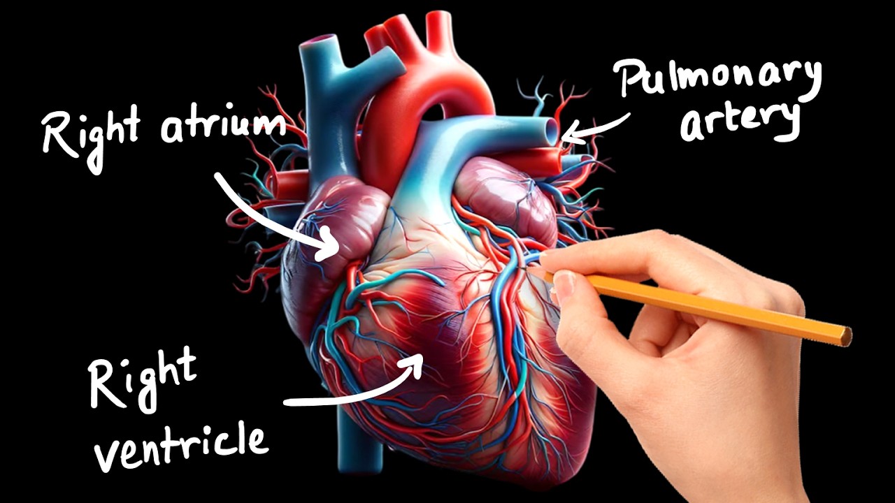
Right atrium receives deoxygenated blood from body via the main veins i.e. superior and inferior
vena cavae. When right atrium contracts it passes the deoxygenated blood to right ventricle. The
opening between right atrium and right ventricle is guarded by a valve known as tricuspid valve
(because it has 3 flaps). When right ventricle contracts, the blood is passed to pulmonary trunk,
which carries blood to lungs. Tricuspid valve prevents the backflow of blood from right ventricle
to right atrium. At the base of pulmonary trunk, pulmonary semilunar valve is present which
prevents the backflow of blood from pulmonary trunk to right ventricle.
The walls of left ventricle are the thickest one. These are about a half-inch thick. They have enough force
to push blood into the body. This gives an evidence that the structures of the parts of heart are adaptive
to their functions.
The oxygenated blood from lungs is brought by pulmonary veins to left atrium. Left atrium
contracts and pumps this blood to left ventricle. The opening between left atrium and left ventricle
is guarded by a valve known as bicuspid valve (because it has two flaps). When left ventricle
contracts, it pumps the oxygenated blood in aorta, which carries blood to all parts of body (except
lungs). Bicuspid valve prevents the backflow of blood from left ventricle to left atrium. At the base
of aorta, aortic semilunar valve is present which prevents the backflow of blood from aorta to
left ventricle (Figure 9.15).
#humanheart #humanheartinhindi
#visiblescience
#alevelbiology
#mdcatbiology
Повторяем попытку...
Доступные форматы для скачивания:
Скачать видео
-
Информация по загрузке:


![Anatomy of the Heart: Structures and Blood Flow [Cardiology Made Easy]](https://imager.clipsaver.ru/1b4V09HzhBw/max.jpg)