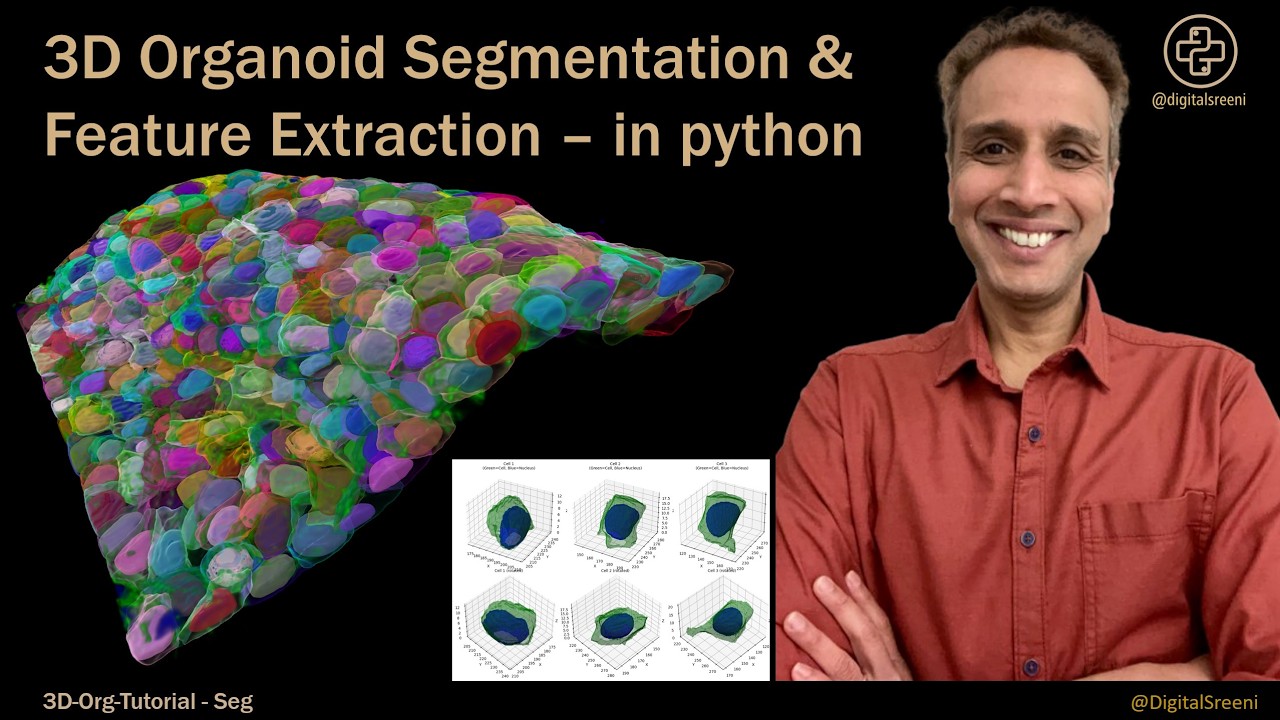
3D Organoid Segmentation and Feature Extraction - in python
Автор: DigitalSreeni
Загружено: 2026-02-20
Просмотров: 342
Описание:
Learn how to segment cells and nuclei in 3D organoid images using Cellpose, perform quality control, and extract quantitative features ready for analysis. This tutorial bridges the gap between raw microscopy data and the structured feature tables we use in subsequent analysis tutorials.
In this tutorial, you'll learn how to take raw 3D fluorescence microscopy images and transform them into analyzable data through automated segmentation and feature extraction using Python.
We cover the complete pipeline from raw TIFF stacks to CSV feature tables. You'll segment nuclei and cells in 3D using Cellpose deep learning models, match each nucleus to its parent cell, apply quality control filters to remove artifacts, and extract morphological, intensity, topology, and shape features per cell. By the end, you'll have production-ready code for analyzing your own organoid datasets.
DATASET:
PDAC organoid images from Ong et al., Nature Methods 2025
Download: https://github.com/quantacell/3DcellS...
Files needed: PDAC-C1.tif (nuclei channel), PDAC-C2.tif (cell channel)
CODE:
Link to complete notebook: https://github.com/bnsreenu/3D-Organo...
COMMERCIAL ALTERNATIVE:
If you're in biopharma and need to scale this workflow to hundreds of organoids across multi-well plates without coding, check out ZEISS arivis. It provides automated segmentation, batch processing, and 3D visualization designed for high-throughput organoid screening.
Повторяем попытку...
Доступные форматы для скачивания:
Скачать видео
-
Информация по загрузке:


![Что ошибочно пишут в книгах об ИИ [Двойной спуск]](https://imager.clipsaver.ru/z64a7USuGX0/max.jpg)