Cranial Nerve Anatomy - The extra-oculor motor nerves (III, IV, VI) (part 1: anatomy)
Автор: The Neuroradiologist
Загружено: 2025-04-10
Просмотров: 5814
Описание:
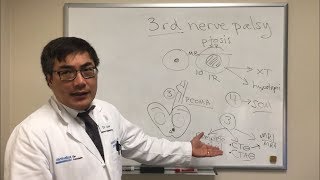
Imaging in diplopia: what to double check in a patient with double vision! In this video, I discuss the anatomy of the extra-ocular motor nerves (oculomotor, trochlear, and abducens nerve) and illustrate their functions and anatomy, with clear correlations to how these structures impact eye movement and coordination. This video is part one of a two-part series, with the second part covering pathology. This video is aimed at medical students, radiology residents, and ophthalmology residents and will enhance your understanding of the imaging anatomy of these vital nerves.
0:00 - Introduction, what is diplopia.
3:56 - Topics
4:27 - Part 1: function
8:05 - Abducens palsy
8:47 - Trochlear palsy
9:43 - Oculomotor palsy
11:05 - Denervation patterns
13:37 - Part 2: Imaging Anatomy
13:46 - Extra-oculor motor nerve segments
15:11 - Brainstem segments
18:17 - Cisternal segments
22:39 - Dorello's canal (abducens nerve)
25:54 - Cavernous Sinus Anatomy
30:48 - Oculomotor cistern
32:05 - Extracranial segments
#neuroradiology #neurology #ophthalmology #brainanatomy #diplopia #medicaleducation #medical #radiology #medicalstudent #cranialnerves #neuroanatomy
Повторяем попытку...

Доступные форматы для скачивания:
Скачать видео
-
Информация по загрузке: