Fungal elements in KOH mount microscopy
Автор: MedLabSolutions
Загружено: 2024-06-25
Просмотров: 350
Описание:
Potassium hydroxide (KOH) mount microscopy is a simple and widely used technique in clinical laboratories to detect fungal elements in clinical specimens. The KOH mount is particularly useful for identifying dermatophytes, yeasts, and other fungi directly from skin, hair, nail, and other tissue samples. This method helps in the rapid diagnosis of fungal infections, enabling timely and appropriate treatment.
Principle of KOH Mount
KOH mount works on the principle that KOH dissolves keratin and other cellular material, which are common components of human tissues, without affecting the rigid cell walls of fungi. This selective dissolution clears the specimen, making fungal elements more visible under the microscope. Typically, a 10-20% KOH solution is used, sometimes combined with dimethyl sulfoxide (DMSO) to enhance the clearing process.
Procedure
Specimen Collection: Samples such as skin scrapings, nail clippings, hair pluckings, or tissue biopsies are collected from the infected area.
Preparation: The specimen is placed on a clean glass slide, and a drop of KOH solution is added.
Cover Slip Application: A cover slip is placed over the specimen to avoid air bubbles and ensure even distribution of the solution.
Incubation: The slide is gently heated or left at room temperature for several minutes to allow the KOH to act on the sample.
Microscopy: The slide is examined under a light microscope using low and high power magnification to identify fungal elements.
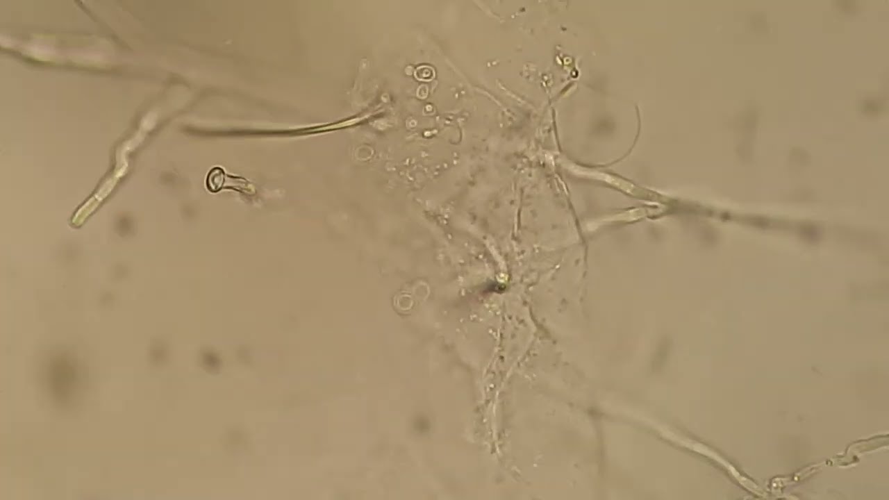
Fungal Elements Identified in KOH Mount
Several fungal structures can be identified in a KOH mount, including:
Hyphae: Long, branching filamentous structures that are characteristic of molds. They can be septate (having cross-walls) or non-septate.
Pseudohyphae: Chains of elongated yeast cells that resemble hyphae but are constricted at the septa. Commonly seen in Candida species.
Spores and Conidia: Reproductive structures of fungi. Conidia are asexual spores produced by molds like Aspergillus.
Budding Yeasts: Round to oval yeast cells that reproduce by budding. Typical of Candida and Cryptococcus species.
Arthroconidia: Spores formed by the fragmentation of hyphae, seen in fungi like Coccidioides.
Chlamydospores: Thick-walled resting spores seen in Candida albicans.
Common Fungi Detected by KOH Mount
Dermatophytes: Fungi causing skin, hair, and nail infections (e.g., Trichophyton, Microsporum, Epidermophyton).
Candida Species: Yeasts causing mucocutaneous and systemic infections.
Aspergillus: Molds causing respiratory infections and invasive aspergillosis.
Cryptococcus: Yeasts causing meningitis and pulmonary infections, particularly in immunocompromised patients.
Malassezia: Yeasts causing tinea versicolor and seborrheic dermatitis.
Coccidioides: Dimorphic fungi causing coccidioidomycosis.
Clinical Significance
KOH mount microscopy is a rapid, cost-effective, and relatively simple diagnostic tool. It is highly useful in the following clinical scenarios:
Dermatophytosis: Identifying dermatophyte infections of the skin, hair, and nails.
Onychomycosis: Detecting fungal infections of the nails.
Candidiasis: Diagnosing mucocutaneous and invasive Candida infections.
Pulmonary Mycoses: Identifying fungal elements in respiratory specimens.
Cutaneous Mycoses: Diagnosing superficial fungal infections.
Advantages and Limitations
Advantages:
Rapid and inexpensive.
Simple to perform with minimal equipment.
Effective for detecting a wide range of fungal elements.
Limitations:
May not differentiate between different fungal species.
Requires experience to interpret results accurately.
False negatives can occur if the fungal load is low or specimen quality is poor.
Conclusion
KOH mount microscopy remains a cornerstone technique in the diagnosis of fungal infections. Its ability to quickly and efficiently reveal fungal elements directly from clinical specimens makes it invaluable in both dermatological and general clinical practice. Proper technique and experience in interpretation are essential to maximize its diagnostic potential.
Повторяем попытку...
Доступные форматы для скачивания:
Скачать видео
-
Информация по загрузке:


