MRT (Magnetic Resonance Tomography) - left knee (MRI/IRM)
Автор: maxtc
Загружено: 2016-06-19
Просмотров: 1176
Описание:
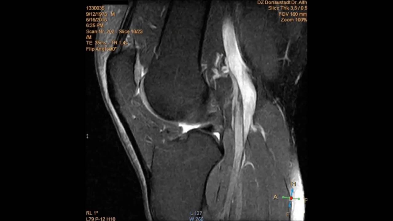
MRT/MRI of my left knee with the typical sound. (Philips Intera 1,5 Tesla)
--------------------------------------------------------------------------------------------------------------------------------------------
MRT Untersuchung meines linken Knies. MRT-Gerät: Philips Intera 1,5 Tesla
--------------------------------------------------------------------------------------------------------------------------------------------
Die Magnetresonanztomographie MRT, kurz auch MR, (Tomographie von altgriechisch τομή tome ‚Schnitt‘ und γράφειν graphein ‚schreiben‘) ist ein bildgebendes Verfahren, das vor allem in der medizinischen Diagnostik zur Darstellung von Struktur und Funktion der Gewebe und Organe im Körper eingesetzt wird. Es basiert physikalisch auf den Prinzipien der Kernspinresonanz (engl. Nuclear Magnetic Resonance, NMR), insbesondere der Feldgradienten-NMR, und wird daher auch als Kernspintomographie bezeichnet (umgangssprachlich gelegentlich zu Kernspin verkürzt). Die ebenfalls zu findende Abkürzung MRI stammt von der englischen Bezeichnung Magnetic Resonance Imaging.
Im Gerät wird keine belastende Röntgenstrahlung oder andere ionisierende Strahlung erzeugt oder genutzt. Allerdings sind die Wirkungen der magnetischen Wechselfelder auf lebendes Gewebe nicht vollständig erforscht.
--------------------------------------------------------------------------------------------------------------------------------------------
Una imagen por resonancia magnética (IRM), también conocida como tomografía por resonancia magnética (TRM) o imagen por resonancia magnética nuclear (IRMN, o NMRI por sus siglas en inglés Nuclear Magnetic Resonance Imaging) es una técnica no invasiva que utiliza el fenómeno de la resonancia magnética nuclear para obtener información sobre la estructura y composición del cuerpo a analizar. Esta información es procesada por ordenadores y transformada en imágenes del interior de lo que se ha analizado.
Es utilizada principalmente en medicina para observar alteraciones en los tejidos y detectar cáncer y otras patologías. También es utilizada industrialmente para analizar la estructura de materiales tanto orgánicos como inorgánicos.
La IRM no debe ser confundida con la espectroscopia de resonancia magnética nuclear, una técnica usada en química que utiliza el mismo principio de la resonancia magnética para obtener información sobre la composición de los materiales.
A diferencia de la TC, no usa radiación ionizante, sino campos magnéticos para alinear la magnetización nuclear de (usualmente) núcleos de hidrógeno del agua en el cuerpo.
--------------------------------------------------------------------------------------------------------------------------------------------
Magnetic resonance imaging (MRI), nuclear magnetic resonance imaging (NMRI), or magnetic resonance tomography (MRT) is a medical imaging technique used in radiology to image the anatomy and the physiological processes of the body in both health and disease. MRI scanners use strong magnetic fields, radio waves, and field gradients to form images of the body.
MRI is based upon the science of Nuclear Magnetic Resonance (NMR). Certain atomic nuclei can absorb and emit radio frequency energy when placed in an external magnetic field. In clinical and research MRI, hydrogen atoms are most-often used to generate a detectable radio-frequency signal that is received by antennas in close proximity to the anatomy being examined. Hydrogen atoms exist naturally in people and other biological organisms in abundance, particularly in water and fat. For this reason, most MRI scans essentially map the location of water and fat in the body. Pulses of radio waves are used to excite the nuclear spin energy transition, and magnetic field gradients localize the signal in space. By varying the parameters of the pulse sequence, different contrasts can be generated between tissues based on the relaxation properties of the hydrogen atoms therein. Since its early development in the 1970s and 1980s, MRI has proven to be a highly versatile imaging modality. While MRI is most prominently used in diagnostic medicine and biomedical research, it can also be used to form images of non-living objects. MRI scans are capable of producing a variety of chemical and physical data, in addition to detailed spatial images.
MRI is widely used in hospitals and clinics for medical diagnosis, staging of disease and follow-up without exposing the body to ionizing radiation.
Повторяем попытку...
Доступные форматы для скачивания:
Скачать видео
-
Информация по загрузке: