Observing the galleries of the olive fruit fly Bactrocera oleae inside the olive using micro-CT
Автор: Javier Alba-Tercedor
Загружено: 2024-10-04
Просмотров: 252
Описание:
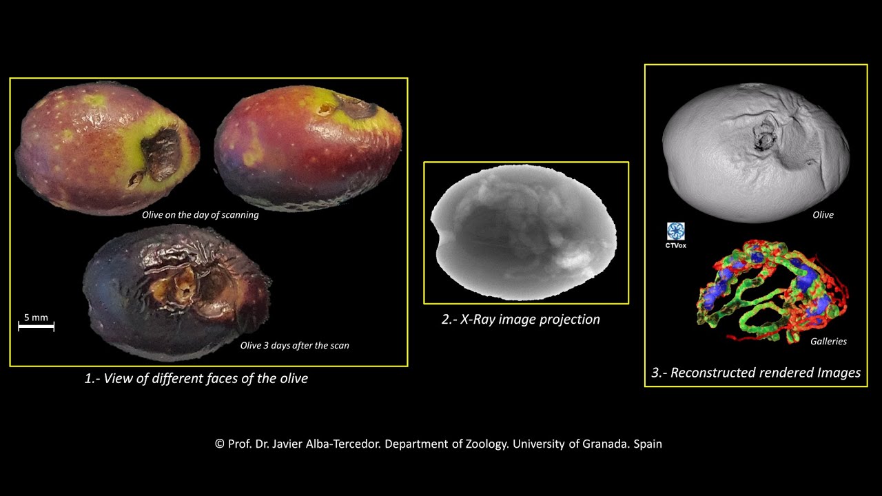
Here is a journey inside an olive fruit attacked by the olive fruit fly in its summer generation, in which can be observed from the beginning at the point of insertion of the ovipositor for laying, as the larvae have been advancing making galleries of increasing thickness as they change from instar 1 to 3. Microtomography has allowed us to reconstruct the galleries in successive colors (red, green, and blue) according to the light of the galleries, corresponding to the three larval instars. Also, near the surface in the vicinity of two adult exit holes, the remains of the puparia can be observed.
The olive was scanned at the Zoology Department of the University of Granada with a Skyscan 1172 microtomograph, using a 0.5mm Aluminium filter and the following scanning parameters: Voxel size= 13.54 µm3, 48 kV, 124 µA, 2x2 camera binning, 180º rotation and a rotation step=0.47°.
Skyscan's CTAnalyser software (v. 1.20.8.0) was used for preliminary cleaning and image segmentation, followed either by Skyscan's Ctvox (v. 3.3.1) or FEI's Amira software (v. 2019.3) for renderings and video recording.
PowerPoint 2013 was used to compose, edit and record the final movie.
The professor of the University of Granada, Francisca Ruano Diaz who is currently researching on the olive pest (within her studies of applied entomology) encouraged to carry out this microtomographic study,
For more details see the paper:
ALBA-TERCEDOR, J., & RUANO, F. (2024). Use of micro-computed tomography to monitor olive fruit damage caused by three insect pests. Scientific Reports, 14(21067):1-10 https://doi.org/10.1038/s41598-024-72...
Повторяем попытку...
Доступные форматы для скачивания:
Скачать видео
-
Информация по загрузке:


