Pseudokeratoconus! Multiple “coincidences” and unraveling this complex case: Kanellopoulos, MD
Автор: LaserVision Ambulatory Eye Surgery Unit, Greece
Загружено: 2022-03-30
Просмотров: 305
Описание:
Let’s travel through this very interesting case that underlines the importance of careful correlation of advanced Cornial diagnostics, slip-lamp biomicroscopy and critical clinical thinking 🤔 ; not everything is really what it appears to be !!!
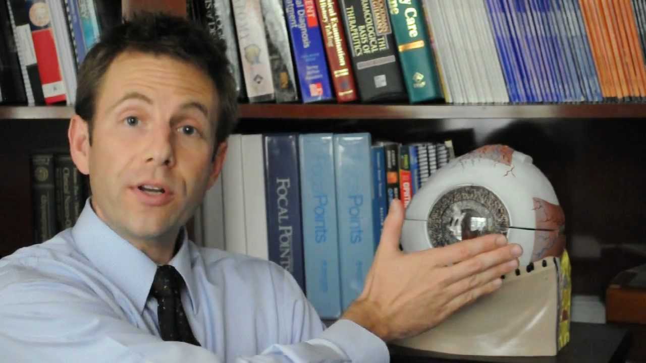
Step by step assessment of each one of the two eyes unravel the fascinating story behind this case that was labeled as advancing keratoconus in a 63-year-old male who is seeking treatment for his poor vision in his right eye.
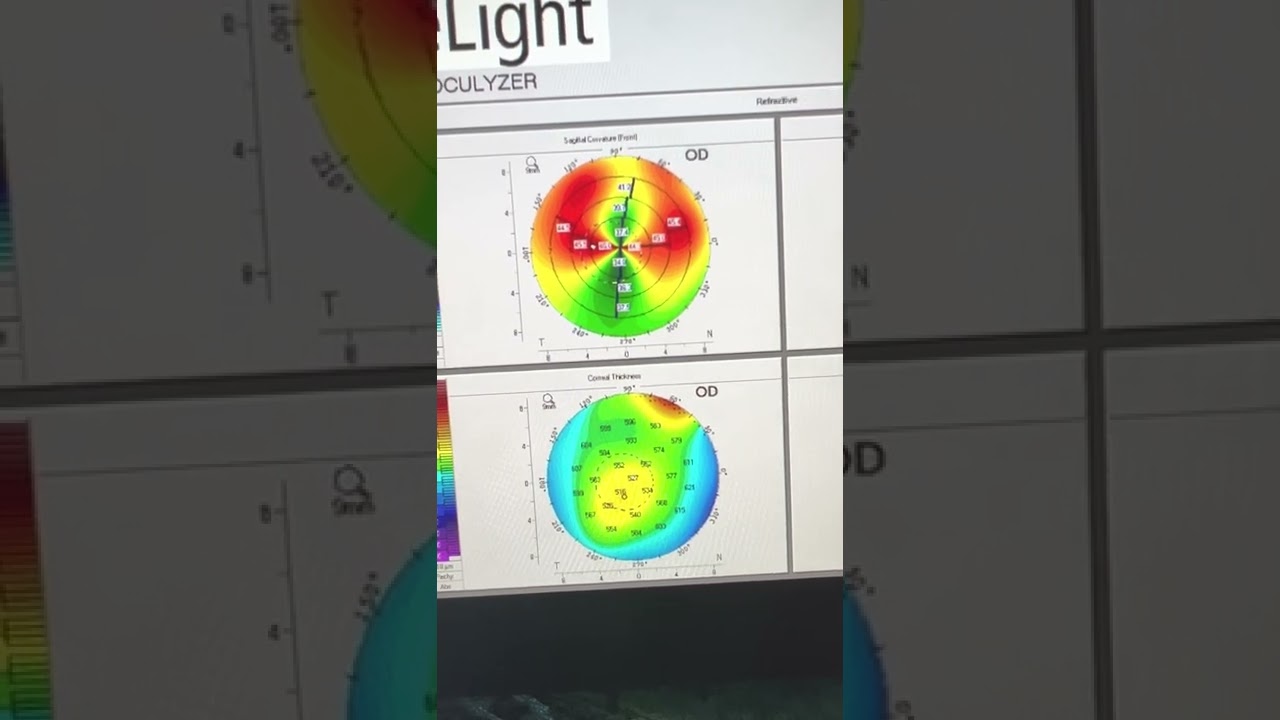
Scheimpflug Corneal tomography, anterior segment OCT corneal mapping and epithelial mapping by the Optovue Avanti Angio-flow, plus careful clinical evaluation on the slit lamp and past sye history, will unravel this complex case. The tjinner due to keratoconus is actually the left eye that has 20/20 vision but due to some past metal foreign body corneal injuries - our patient was a craftsman in his earlier years-has developed central flattening and thus very good visual acuity from this acquired by accidental “intervention” .
The right eye that came to us with a diagnosis of keratoconus, has actually the milder keratoconus-related thinning has developed severe against-the-rule astigmatism, resembling kindof “upside down” pellucid marginal degeneration due to a unrelated superior corneal marginal degeneration-a separate phenomena or pathology if you may- to the underlying keratoconus, and as a result uncorrected 20/200 vision and best corrected 20/40-
Of all the ectasia and suspect cases that I have evaluated I think this single case summarizes the importance of careful correlation between corneal imaging, slit-lamp biomicroscopy and clinical judgment. The appropriate treatment plan and options are also discussed
Повторяем попытку...
Доступные форматы для скачивания:
Скачать видео
-
Информация по загрузке: