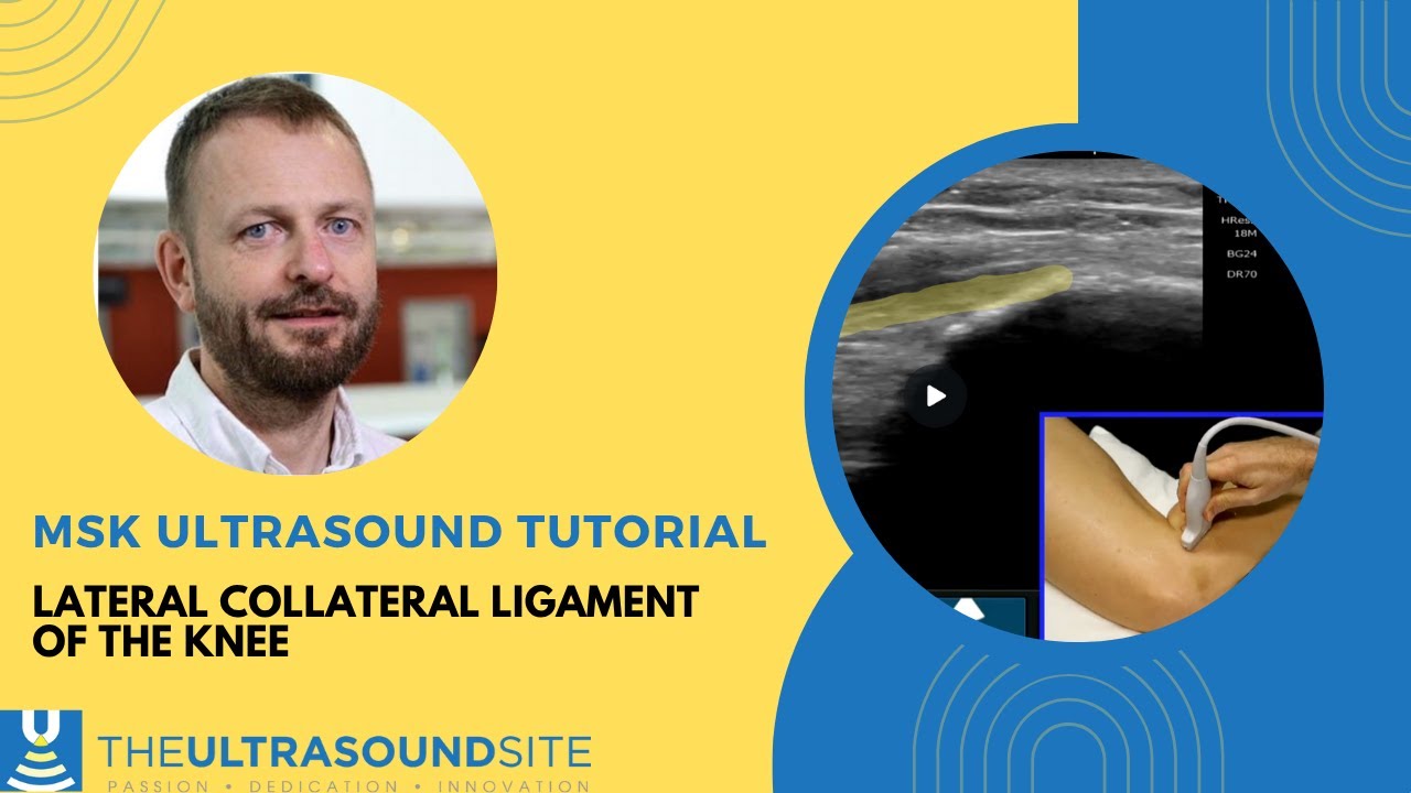
Lateral Knee MSK Ultrasound Targeted Scanning Demo of LCL and Biceps Femoris Insertion on the Fibula
Автор: Learn MSK Sono
Загружено: 2023-07-27
Просмотров: 1117
Описание:
Most practitioners enjoy scanning the knee because the course of the tendons and ligaments are relatively straight and easy to scan compared to the other joints of the extremities. If there was a tricky part of the knee to scan, this is it! I Hope my demo shortens the learning curve for you!
Points to remember:
✔️ The Lateral Meniscus is your home base
to start your examination of the lateral
knee due to its easily recognizable
appearance.
✔️ From the Lateral Meniscus and Tibia,
slide your probe posterior and inferior (while staying at the same angle) until the Fibula comes into view.
✔️ Both the LCL and Biceps Femoris Tendon insert onto the Fibula.
✔️ Keep the distal portion of your probe
fixed on the Fibula to image both structures.
✔️ Rotate the proximal portion of your probe
anteriorly to isolate the LCL.
✔️ Rotate the proximal portion of your probe
posteriorly to isolate the Biceps Femoris.
Повторяем попытку...

Доступные форматы для скачивания:
Скачать видео
-
Информация по загрузке: