BODY CAVITIES ANATOMY - Cranial, Spinal, Thoracic, Abdominopelvic
Автор: Neural Academy
Загружено: 2024-01-02
Просмотров: 16649
Описание:
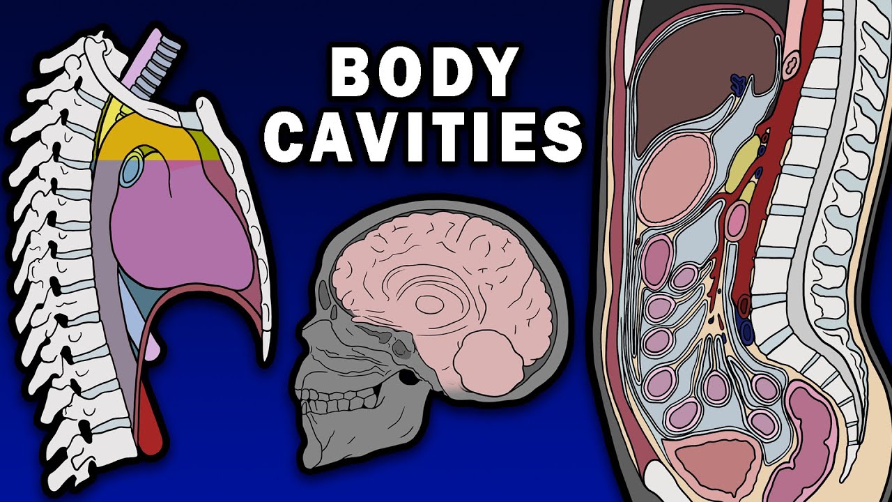
The internal organs are inside body cavities. The 2 main ones are the dorsal and ventral cavities. The dorsal cavity includes the cranial cavity, which houses the brain, and the spinal canal, which houses the spinal cord.
The ventral cavity contains the thoracic and abdominopelvic cavities. The thoracic cavity contains the mediastinum and pleural cavities. The mediastinum contains the heart, trachea, and esophagus.
The mediastinum has 2 divisions – superior and inferior, which can be subdivided into anterior, middle, and posterior divisions.
The pleural cavities contain the lungs.
Found beneath the diaphragm, the abdominopelvic cavity can be divided into the abdominal and pelvic cavities. Since there is no actual membrane that separates the abdominal cavity from the pelvic cavity, sometimes this area is called the abdominal pelvis or the peritoneal cavity.
The peritoneal cavity is the potential space between the parietal and visceral peritoneum. The peritoneum is a serous membrane lining the viscera and the abdominal cavity wall. The parietal peritoneum lines the internal surface of the abdominopelvic wall. The visceral peritoneum wraps around the organs. The omentum, a layer of sheets of visceral peritoneum, extends from the stomach and proximal part of the duodenum and covers the front of your abdomen like an apron, while the mesentery, a double layer in the back, attaches the intestines to the back abdominal wall.
The greater omentum is made up of four sheets of visceral peritoneum. Descending from the stomach’s greater curvature and the proximal part of the duodenum, it folds and then attaches to the anterior surface of the transverse colon.
The lesser omentum, a double layer of visceral peritoneum, attaches the lesser curvature of the stomach and the proximal part of the duodenum to the liver. It is made up of a flat sheet called the hepatogastric ligament, as well as the hepatoduodenal ligament’s free edge, which contains the portal triad.
A peritoneal ligament is a double fold of peritoneum connecting either viscera to other viscera or viscera to the abdominal wall. Splenic peritoneal ligaments include the phrenicocolic, gastrosplenic, and splenorenal ligaments. Gastric ligaments include the gastrophrenic and gastrocolic ligaments. And hepatic ligaments include the falciform, hepatogastric, and hepatoduodenal ligaments.
The peritoneal cavity can be subdivided into the greater and lesser peritoneal sacs. The greater sac is divided yet further by the mesentery of the transverse mesocolon into the supracolic compartment anterior and superior to the transverse mesocolon, and the infracolic compartment posterior and inferior to the transverse mesocolon. These are connected by the paracolic gutters. The supracolic compartment contains the liver, spleen, and stomach. The infracolic compartment contains the ascending and descending colon and the small intestine. The mesentery of the small intestine divides the infracolic compartment into left and right infracolic spaces.
The lesser sac is found behind the liver and stomach in front of the pancreas and duodenum. It is needed to provide a space for the stomach to move unhindered. It is connected to the greater sac through the epiploic, or omental, foramen of Winslow, an opening in the omental bursa.
The peritoneal cavity is different in structure in males and females. The peritoneal cavity is completely sealed in males, but not in females. Males have a rectovesical pouch, a double folding of peritoneum, found between the rectum and bladder. Females have the rectouterine pouch, a double folding of the peritoneum between the rectum and the back wall of the uterus. They also have a vesicouterine pouch, which is another double folding of peritoneum between the front of the uterus and the bladder. The fallopian tubes open into the peritoneal cavity, so the female genital tract is technically connected to the abdominal cavity.
Abdominal viscera can be grouped anatomically based on their relationship to the peritoneum into intraperitoneal and retroperitoneal organs. Intraperitoneal organs are enveloped, both anteriorly and posteriorly, by the visceral peritoneum. These include the stomach, liver, spleen, parts of the duodenum, the jejunum, ileum, transverse colon, and sigmoid colon.
Meanwhile, retroperitoneal organs are not associated with visceral peritoneum. They're only being covered by parietal peritoneum on their anterior surface. Here’s a mnemonic to remember which abdominal viscera are retroperitoneal: SAD PUCKER
Retroperitoneal organs can be further subdivided into primarily retroperitoneal and secondarily retroperitoneal organs. Primarily retroperitoneal organs developed and stayed outside the parietal peritoneum, while secondarily retroperitoneal organs were initially intraperitoneal, suspended by mesentery, and then became retroperitoneal when the mesentery fused with the posterior abdominal wall (only their anterior surface is covered with peritoneum).
Повторяем попытку...
Доступные форматы для скачивания:
Скачать видео
-
Информация по загрузке:


![Body Cavities and Membranes: Drawn and Defined [Anatomy Physiology]](https://image.4k-video.ru/id-video/sqEDTVYq1_U)


