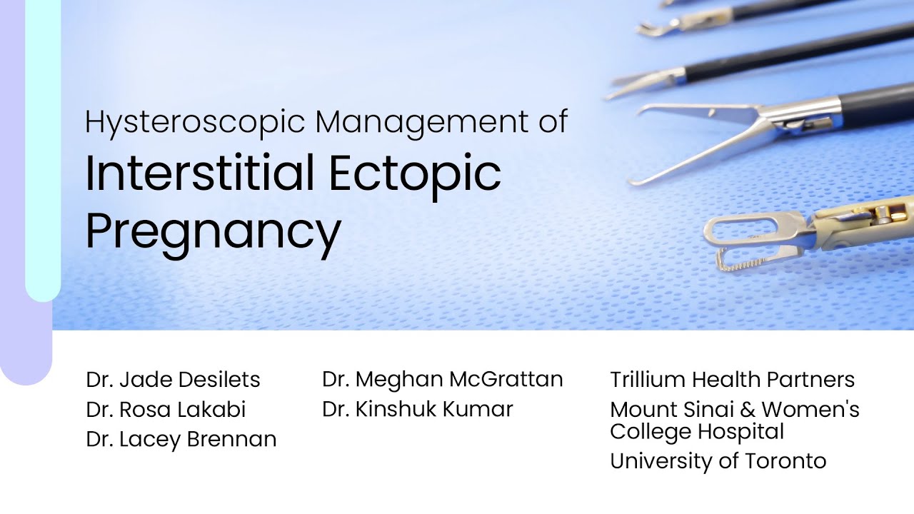
Hysteroscopic Management of Interstitial Ectopic Pregnancy
Автор: CanSAGE Secretariat
Загружено: 2025-09-22
Просмотров: 272
Описание:
This video demonstrates the hysteroscopic management of a proximal interstitial ectopic pregnancy, outlining key diagnostic features, laparoscopic confirmation, and a minimally invasive stepwise resection technique that preserves uterine integrity.
See full transcript here: https://cansagevideos.com/hysteroscopic-ma...
Presented By
Dr. Jade Desilets
Dr. Rosa Lakabi
Dr. Lacey Brennan
Dr. Meghan McGrattan
Dr. Kinshuk Kumar
Affiliations
Trillium Health Partners
Mount Sinai & Women’s College Hospital
University of Toronto
This instructional video presents the hysteroscopic treatment of a proximal interstitial ectopic pregnancy—a rare but potentially life-threatening form of ectopic implantation occurring within the intramural segment of the fallopian tube. The patient, a 38-year-old G3P0, presented with persistent spotting two months after a dilation and curettage. A thin endometrium and a 2 cm fundal lesion with minimal myometrial thickness were visualized on ultrasound, and a sonohysterogram confirmed a lesion communicating with the uterine cavity. These findings were consistent with a proximal interstitial ectopic pregnancy.
Interstitial ectopic pregnancies account for less than 3% of all ectopic gestations but carry a markedly higher risk of hemorrhage and mortality if ruptured. Diagnostic ultrasound criteria include an empty uterine cavity, a gestational sac located at least 1 cm from the lateral uterine edge, a surrounding myometrial thickness of ≤5 mm, and the characteristic interstitial line sign connecting the sac to the endometrial canal. Accurate localization is essential to distinguish interstitial from cornual or intrauterine pregnancies, as management strategies differ significantly.
Laparoscopy confirmed a proximal interstitial implantation with lateral fundal distension and medial displacement of the round ligament. The procedure followed a structured four-step approach: (1) laparoscopic and hysteroscopic confirmation of location, (2) vasopressin injection to reduce bleeding, (3) hysteroscopic resection under laparoscopic guidance using a 6 mm hysteroscope and mechanical tissue removal system, and (4) tubal dye testing to confirm patency and assess the resection site. Real-time laparoscopic transillumination aided in monitoring residual myometrial thickness and guiding precise tissue removal.
Final hysteroscopic inspection confirmed an empty left cornua with intact myometrium. Postoperatively, the patient’s beta-hCG normalized, menses resumed, and follow-up hysterosonogram showed a normal uterine cavity. This case underscores the value of combined hysteroscopic and laparoscopic visualization for accurate diagnosis and safe treatment of proximal interstitial ectopic pregnancies. When performed in appropriately selected cases, hysteroscopic management provides a fertility-preserving, minimally invasive alternative to cornuectomy or salpingectomy.
Повторяем попытку...
Доступные форматы для скачивания:
Скачать видео
-
Информация по загрузке: