Pulmonary Hypertension in a dog
Автор: Kahina Kartout
Загружено: 2023-05-26
Просмотров: 1692
Описание:
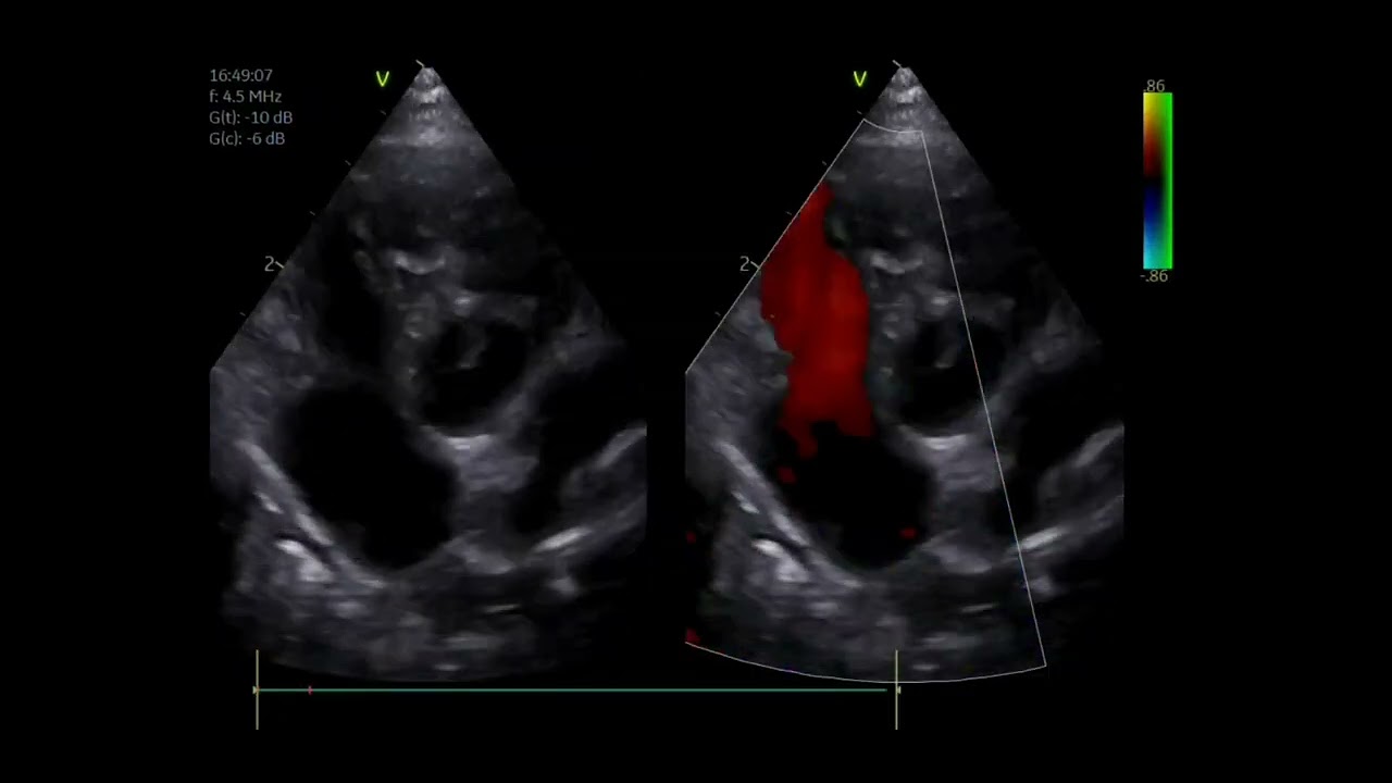
PA pressure can be estimated noninvasively using continuous wave (CW) Doppler echocardiography when TR or pulmonary valve regurgitation (PR) is present and when pulmonic stenosis (or other RV outflow obstruction) is absent.
Color Doppler imaging guides CW Doppler cursor placement, and might identify a measurable tricuspid (or pulmonary) regurgitant jet when an audible murmur is absent.
Injection of agitated saline can sometimes improve the signal strength of the regurgitant jet.
With TR, the modified Bernoulli relationship is used to estimate the systolic pressure gradient between the right ventricle and its atrium, based on the highest TR jet velocity that can be recorded. This gradient is then added to the estimated RA pressure.
It has been recommended that PH in dogs be defined as “an intermediate or high probability of PH” when characteristic echo findings are identified in animals with compatible clinical signs. For dogs (and presumably cats and horses), this includes a TR peak velocity more than 3.4 m/s in cases with measurable TR and without RV outflow obstruction.
Typical 2D and M-mode echo findings consistent with PH include: RV chamber dilation, some degree of RV hypertrophy (wall thickening), abnormal septal motion, and PA dilation .
Impaired RV function, RA enlargement and caudal vena caval distension also might be found.
In one series of dogs with PH, about half had normal RV size and wall thickness and less than a third had severe RV dilation; RV hypertrophy was considered moderate in less than 25% of cases, and severe in only 10% (Johnson, 1999). Severe RV concentric hypertrophy is most likely to occur in young animal.
RV systolic function can be assessed by measuring the tricuspid annular plane excursion (TAPSE), because RV contraction occurs mostly along the longitudinal plane. Dogs with severe PH are more likely to have a TAPSE below reference range for their body weight. Yet, TAPSE often is normal in dogs with mild or moderate PH, especially those with postcapillary PH (that is, left heart disease), where it might be increased (hyperdynamic) because of septal tethering to the left side. For dogs with DMVD, there was no difference in TAPSE between those with PH and those without (Poser, 2017). This suggests either that RV function is not decreased, or more likely, that the hyperdynamic left ventricle falsely increases TAPSE. Falsely increased TAPSE also can occur with severe TR. Ratios of TAPSE/Ao or adjusted TAPSE/Ao, and weight-adjusted TAPSE could serve as a body weight-independent means to identify severe PH, as well as to assess RV function, even in dogs with DMVD (Caivano, 2018a).
Source 📚 : New edition of Cardiovascular Disease in Companion Animals. Ware Wendy A. , Bonagura John D
Повторяем попытку...
Доступные форматы для скачивания:
Скачать видео
-
Информация по загрузке:


