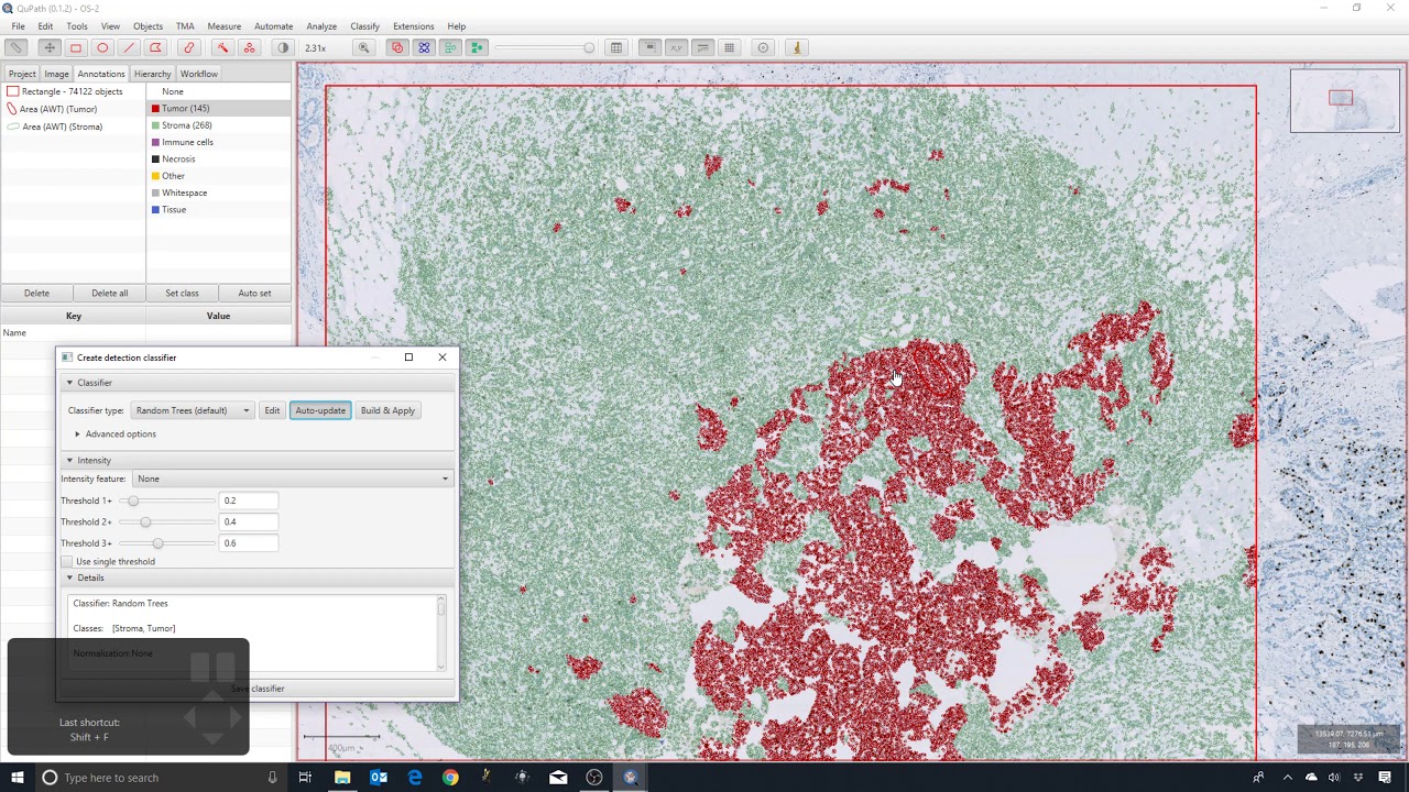
Measuring the Area of DAB in IHC images [QuPath - Thresholding]
Автор: Johanna M. Dela Cruz
Загружено: 2025-01-02
Просмотров: 2037
Описание:
Learn how to use QuPath to threshold regions in IHC images for area measurements. In particular, this tutorial shows how areas stained with DAB are measured. This tutorial is meant for Fiji (ImageJ) users who are looking for an alternative way to measure DAB in their IHC images.
Images used in this tutorial are from the Cell image Library [Larry True, Eric Deutsch, Laura Pascal, Tracy Sherertz, Laura Walashek, David Campbell, Alvin Liu (2012) ]: Tissue sections of human prostate containing adenocarcinoma immunostained for the cell-surface antigen CD90. Bound antibody was detected using avidin-biotin-peroxidase and diaminobenzidine (DAB). Sections were lightly counterstained with hematoxylin (nuclei stained in blue). The images are part of a large collection of images generated from numerous specimens to characterize the distribution of CD90 in human prostate tissue. CIL33962 (doi:10.7295/W9CIL33962), CIL34115 (doi:10.7295/W9CIL34115), CIL34119 (doi:10.7295/W9CIL34119).
SUBSCRIBE to have first access to new video tutorials: / @johanna.m.dela-cruz
Повторяем попытку...
![Measuring the Area of DAB in IHC images [QuPath - Thresholding]](https://imager.clipsaver.ru/828bfTIMvno/max.jpg)
Доступные форматы для скачивания:
Скачать видео
-
Информация по загрузке:


![Measuring Protein Expression and Cellular Fluorescence [Mean Gray Value vs Integrated Density]](https://imager.clipsaver.ru/GaRxdBkoqlQ/max.jpg)