Anatomy of Spleen | Gross |External features | blood supply | clinical
Автор: Knowing Anatomy
Загружено: 2020-07-24
Просмотров: 3941
Описание:
The spleen is an organ found in virtually all vertebrates. Similar in structure to a large lymph node, it acts primarily as a blood filter. The word spleen comes from Ancient Greek σπλήν (splḗn)
The spleen plays important roles in regard to red blood cells (erythrocytes) and the immune system. It removes old red blood cells and holds a reserve of blood, which can be valuable in case of hemorrhagic shock, and also recycles iron. As a part of the mononuclear phagocyte system, it metabolizes hemoglobin removed from senescent red blood cells (erythrocytes). The globin portion of hemoglobin is degraded to its constitutive amino acids, and the heme portion is metabolized to bilirubin, which is removed in the liver.
The spleen synthesizes antibodies in its white pulp and removes antibody-coated bacteria and antibody-coated blood cells by way of blood and lymph node circulation. These monocytes, upon moving to injured tissue (such as the heart after myocardial infarction), turn into dendritic cells and macrophages while promoting tissue healing. The spleen is a center of activity of the mononuclear phagocyte system and is analogous to a large lymph node, as its absence causes a predisposition to certain infections.
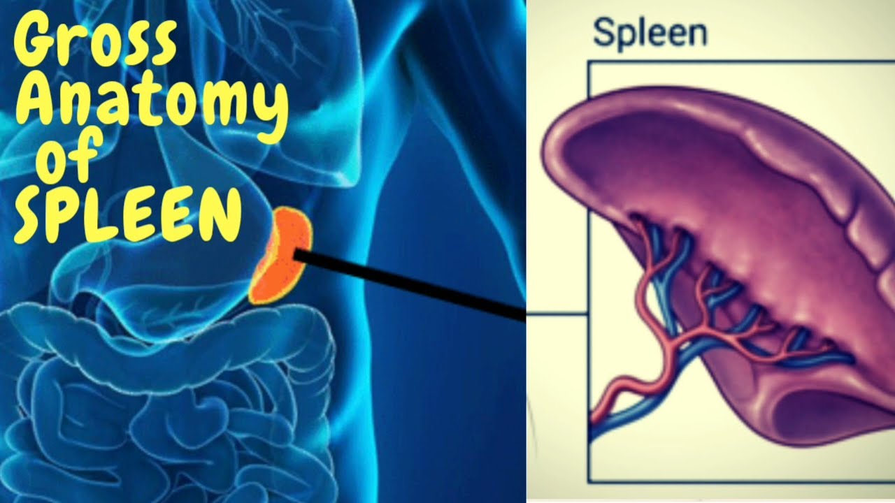
In humans the spleen is purple in color and is in the left upper quadrant of the abdomen.The spleen is underneath the left part of the diaphragm, and has a smooth, convex surface that faces the diaphragm. It is underneath the ninth, tenth, and eleventh ribs. The other side of the spleen is divided by a ridge into two regions: an anterior gastric portion, and a posterior renal portion. The gastric surface is directed forward, upward, and toward the middle, is broad and concave, and is in contact with the posterior wall of the stomach. Below this it is in contact with the tail of the pancreas. The renal surface is directed medialward and downward. It is somewhat flattened, considerably narrower than the gastric surface, and is in relation with the upper part of the anterior surface of the left kidney and occasionally with the left adrenal gland.
Blood supply
Visceral surface of the spleen
Near the middle of the spleen is a long fissure, the hilum, which is the point of attachment for the gastrosplenic ligament and the point of insertion for the splenic artery and splenic vein. There are other openings present for lymphatic vessels and nerves.
Like the thymus, the spleen possesses only efferent lymphatic vessels. The spleen is part of the lymphatic system. Both the short gastric arteries and the splenic artery supply it with blood.
The germinal centers are supplied by arterioles called penicilliary radicles.
Nerve supply
The spleen is innervated by the splenic plexus, which connects a branch of the celiac ganglia to the vagus nerve.
The underlying central nervous processes coordinating the spleen's function seem to be embedded into the Hypothalamic-pituitary-adrenal-axis, and the brainstem, especially the subfornical organ.
Development
The spleen is unique in respect to its development within the gut. While most of the gut organs are endodermally derived (with the exception of the neural-crest derived adrenal gland), the spleen is derived from mesenchymal tissue. Specifically, the spleen forms within, and from, the dorsal mesentery. However, it still shares the same blood supply—the celiac trunk—as the foregut organs.Clinical significance
Surgically removed spleen of a child with thalassemia. It is about 15 times larger than normal.
Thalassemia enlarged spleen after splenectomy
Enlarged spleen
Main article: Splenomegaly
Enlargement of the spleen is known as splenomegaly. It may be caused by sickle cell anemia, sarcoidosis, malaria, bacterial endocarditis, leukemia, pernicious anemia, Gaucher's disease, leishmaniasis, Hodgkin's disease, Banti's disease, hereditary spherocytosis, cysts, glandular fever (mononucleosis or 'Mono' caused by the Epstein–Barr virus), and tumours. Primary tumors of the spleen include hemangiomas and hemangiosarcomas. Marked splenomegaly may result in the spleen occupying a large portion of the left side of the abdomen.
The spleen is the largest collection of lymphoid tissue in the body. It is normally palpable in preterm infants, in 30% of normal, full-term neonates, and in 5% to 10% of infants and toddlers. A spleen easily palpable below the costal margin in any child over the age of 3–4 years should be considered abnormal until proven otherwise.
Повторяем попытку...
Доступные форматы для скачивания:
Скачать видео
-
Информация по загрузке:

![Anatomy of the Heart: Structures and Blood Flow [Cardiology Made Easy]](https://imager.clipsaver.ru/1b4V09HzhBw/max.jpg)