Spotting the Signs: Complex Cervical Mass in Sonography
Автор: Dr. Saima Khan
Загружено: 2025-06-08
Просмотров: 1787
Описание:
Spotting the Signs: Complex Cervical Mass in Sonography #doctor #ultrasound #health @Dr.SaimaKhan
Here's a very detailed ultrasound report based on your provided findings, including differentials and suggested further investigations:
ULTRASOUND PELVIS REPORT
Patient Presentation:
Chief complaints: Severe pelvic pain, foul-smelling and copious vaginal discharge.
Clinical suspicion: Cervical pathology/mass, possible pelvic infection or malignancy.
Ultrasound Findings:
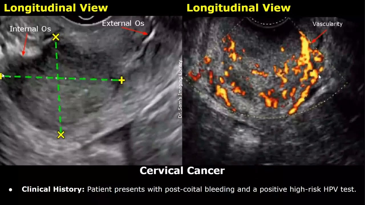
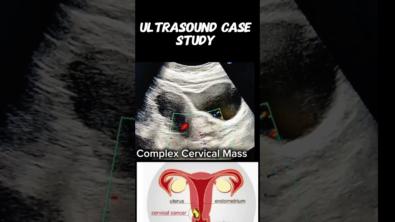
1. Cervical Region:
A well-defined, predominantly hyperechoic heterogeneous mass measuring 64 x 45 mm is noted in the cervical region, abutting the lower uterine segment.
Margins are irregular.
The mass lacks internal vascularity on color Doppler, which may suggest necrosis or a non-viable lesion.
Adjacent cervical stroma appears distorted.
There is no clear cleavage plane between the mass and the lower uterine segment, suggesting possible contiguous involvement.
Associated fluid collection or debris may indicate secondary infection or necrosis.
2. Uterus:
Uterine contour appears normal, endometrial stripe is not well-visualized, likely obscured by adjacent pathology or discharge.
3. Right Ovary:
Contains a simple ovarian cyst with thin walls and anechoic content.
No mural nodules or septations.
4. Left Adnexa:
A complex cystic lesion is noted measuring approximately ___ mm (exact size to be inserted if known), containing a vascular mural nodule.
Internal echoes and septations present.
Doppler reveals increased vascularity within the mural nodule, raising suspicion for neoplastic etiology.
5. Free Fluid:
Trace to mild free fluid noted in the pelvis, with internal echoes, suggesting inflammatory exudate or infected fluid.
Impression:
1. Cervical mass (64 x 45 mm), hyperechoic heterogeneous with irregular margins and no vascularity.
Features may represent:
Cervical carcinoma (necrotic core with poor vascularity)
Infected cervical polyp or prolapsed fibroid with secondary degeneration
Chronic cervical abscess or necrotic infected mass (less likely without systemic signs)
Endocervical stromal tumor (rare)
2. Left adnexal complex cystic lesion with vascular mural nodule:
Differentials:
Borderline ovarian tumor
Ovarian carcinoma
Endometrioma with atypical features
Tubo-ovarian abscess (less likely given the discrete vascular nodule and lack of fever/systemic signs)
3. Right ovarian simple cyst – likely benign.
Recommendations for Further Evaluation:
1. Pelvic MRI with contrast:
To assess soft tissue involvement, vascularity, and extent of cervical and adnexal masses.
Helpful for preoperative planning and staging.
2. Pap smear and HPV typing (if not recently done) – to assess cervical pathology.
3. Cervical biopsy or colposcopic evaluation – to determine histology of cervical mass.
4. CA-125, CEA, HE4, and Risk of Malignancy Index (RMI) – for adnexal mass risk stratification.
5. CBC, CRP, ESR – to assess for infection/inflammation.
6. Diagnostic hysteroscopy or D&C (if cervical canal is patent and infection ruled out) – to assess uterine cavity.
7. Consultation with gynecologic oncology – highly advised due to suspicious nature of lesions.
Urgency Note:
Given the patient's symptoms of severe pelvic pain, malodorous discharge, and complex sonographic findings, urgent evaluation by gynecology and possibly gynecologic oncology is warranted. Infections, if present, should be promptly managed, and malignancy should be ruled out or confirmed through biopsy.
#CervicalMass #PelvicUltrasound #GynecologyImaging #WomensHealth #UltrasoundFindings #RadiologyCase #ComplexMass #CervicalPathology #TransvaginalUltrasound #MedicalImaging #RadiologyEducation #SonographyCase #GynUltrasound #CervicalAnomaly #UltrasoundDiagnosis #PelvicPathology #ImagingMatters #RadiologyLife #DiagnosticUltrasound #ClinicalSonography
YouTube Link:
/ @dr.saimakhan
WhatsApp Channel
whatsapp.com/channel/0029Vaeb2enGJP8OyLo5gp2e
Instagram
instagram.com/saima2269khan/?utm_source=qr&igsh=MXRpcW9oZzlsY3F0dQ==
Facebook
facebook.com/profile.php?id=100004728272539&mibextid=ZbWKwL
Tiktok Account:
https://www.tiktok.com/@drsaimakhan03...
Tags:
#DoctorMike #DrEricBergDC #MedSchoolInsiders #DrDray #ZDoggMD #DrAxe #TheDoctors #DrJoshAxe #InstituteOfHumanAnatomy #MamaDoctorJones #drsaimakhan #dr.saimakhan
Повторяем попытку...
Доступные форматы для скачивания:
Скачать видео
-
Информация по загрузке: