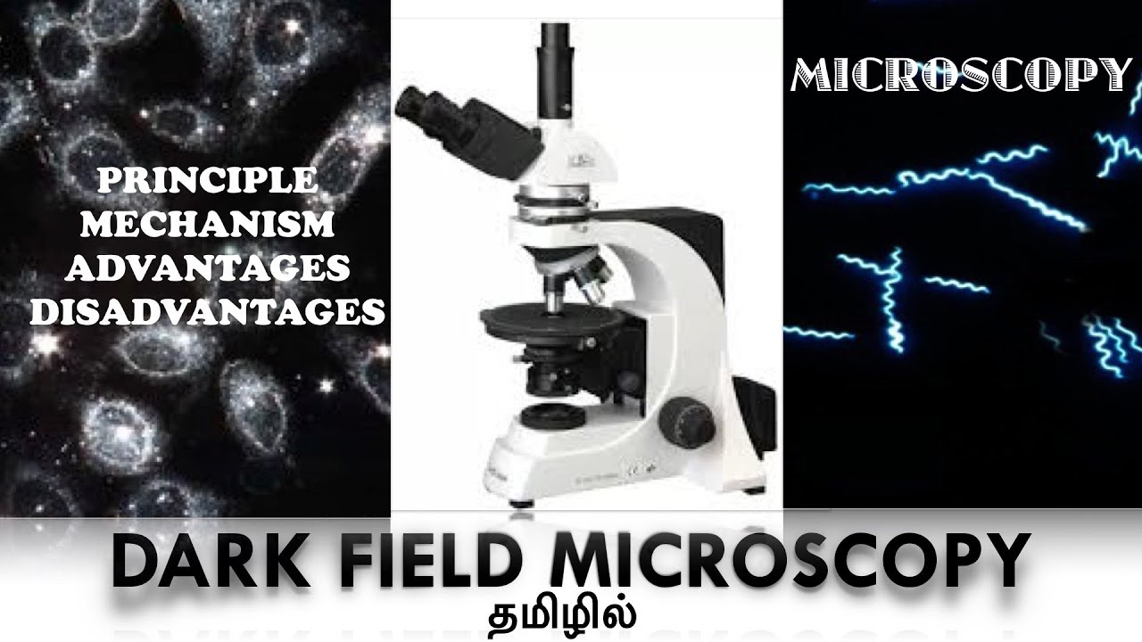
Dark Field Microscopy / Principle / Mechanism / Advantages / Disadvantages / Tamil
Автор: Microbiological Concepts
Загружено: 2020-10-12
Просмотров: 9483
Описание:
In this video, the dark field microscopy, its principle, mechanism, advantages and its disadvantages are discussed.
The dark-field microscope is used to examine detailed images of living, unstained cells and organisms by simply changing the way in which they are illuminated. Instead of the normal condenser, a dark field microscope uses a darkfield condenser that contains an opaque disc.
The disc blocks light that would enter the lens directly, only the reflected light from the specimen enters the objective lens because there is no background light, so the specimen appears bright against the black background.
Principle: The objective and the ocular lenses used in the dark ground microscope are the same as in the ordinary light microscope, however, a special condenser is used, which prevents the transmitted light from directly illuminating the specimen. A hollow cone of light is focused on the specimen in such a way that unreflected and unrefracted rays do not enter the objective. The only light that has been reflected or refracted by the specimen forms an image. The field surrounding a specimen appears black, while the object itself is brightly illuminated.
Working Mechanism: In a dark field microscope, the object is brilliantly illuminated against a dark background. This is accomplished by equipping a light microscope with a special kind of condenser. It is a condenser with a darkfield stop (annular stop), which is an opaque disc (annular filter) obstructing the path of light from the light source centrally but allowing a peripheral ring of light. Thus, the condenser transmits a hollow cone of light from the light source. This cone of light converges on the object and diverges from there again as an inverted hollow cone. Thus, no light enters into the objective, as it remains in the dark cone and the field essentially appears dark in absence of any objects. However, if some objects such as microbial cells are present, some of the light rays are scattered (diffracted) by them. The diffracted rays enter into the objective and reach the eye. Thus, the object (microbial cells) appears bright in a dark microscopic field.
Advantages of Darkfield Microscopy: The dark-field microscope can reveal considerable internal structure in larger eucaryotic microorganisms. It also is used to identify certain bacteria such as the thin and distinctively shaped Treponema pallidum, the causative agent of syphilis. Resolution by dark-field microscopy is somewhat better than bright-field microscopy. Improves image contrast without the use of stain, and thus do not kill cells. Direct detection of non-culturable bacteria present in patient samples. No sample preparation is required. Requires no special set up, even a light microscope can be converted to the dark field.
Disadvantages of Darkfield Microscopy: Necessity to examine wet, moist specimens containing living organisms very quickly because visualization of the moving bacteria is essential to detection.The sample must be very strongly illuminated, which can cause damage to the sample. Besides the sample, dust particles also scatter the light and appear bright. Sample material needs to be spread thinly, dense preparations can grossly affect the contrast and accuracy of the dark field’s image.
If you want to watch the bright field microscopy topic, please click the link below:
• Bright Field Microscopy | Mechanism, Advan...
Повторяем попытку...
Доступные форматы для скачивания:
Скачать видео
-
Информация по загрузке: