Chest X-Ray breakdown: assessing a film with pleural plaques
Автор: theRadiologist
Загружено: 2023-04-22
Просмотров: 16363
Описание:
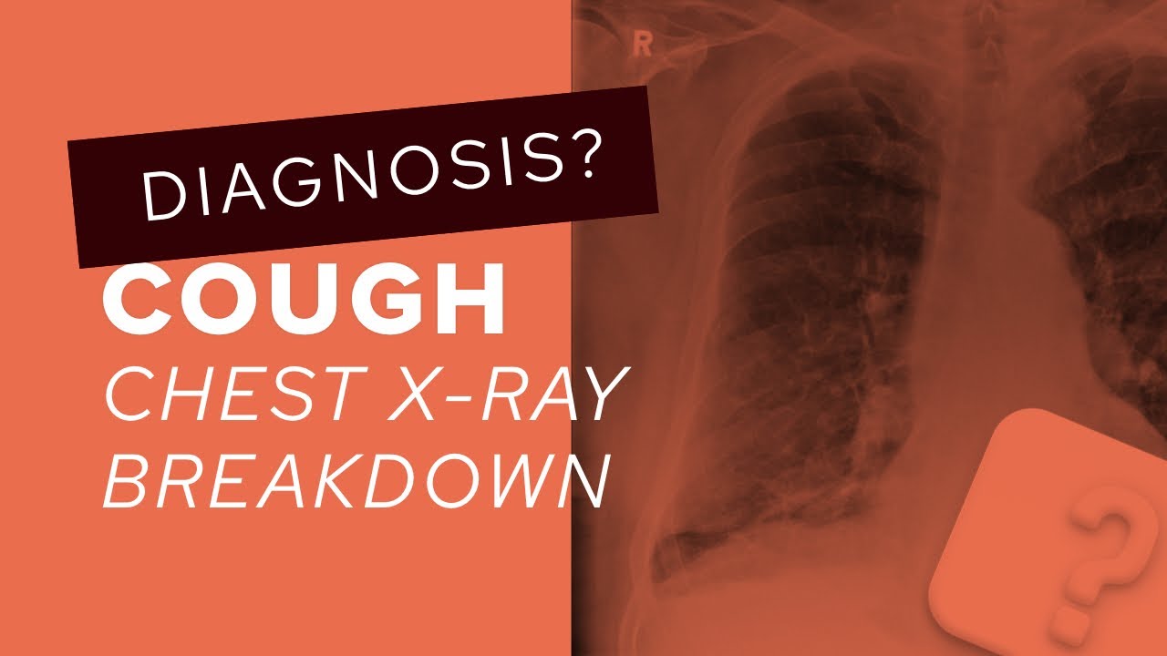
A male in his 70s presents to his GP with a cough. What does the X-Ray show?
————————————————————
LUNG CANCER AND PLEURAL PLAQUES
👨🏽💻Facing a chest X-Ray with a lot going on can be daunting but by going through it systematically you can get to the diagnosis
👨🏽💻There are calcified pleural plaques - irregular high density areas in both lungs giving something called a ‘holly leaf sign’ can point you towards this. If you look at the diaphragm you’ll see bilateral calcified diaphragmatic pleural plaques
👨🏽💻 Pleural plaques are rarely troublesome on their own but point to exposure to asbestos. Asbestos was previously widely used in insulation before it was realised that it was carcinogenic. Asbestos exposure has several manifestations in the lung
▫️CALCIFIED ‘PLEURAL PLAQUES’
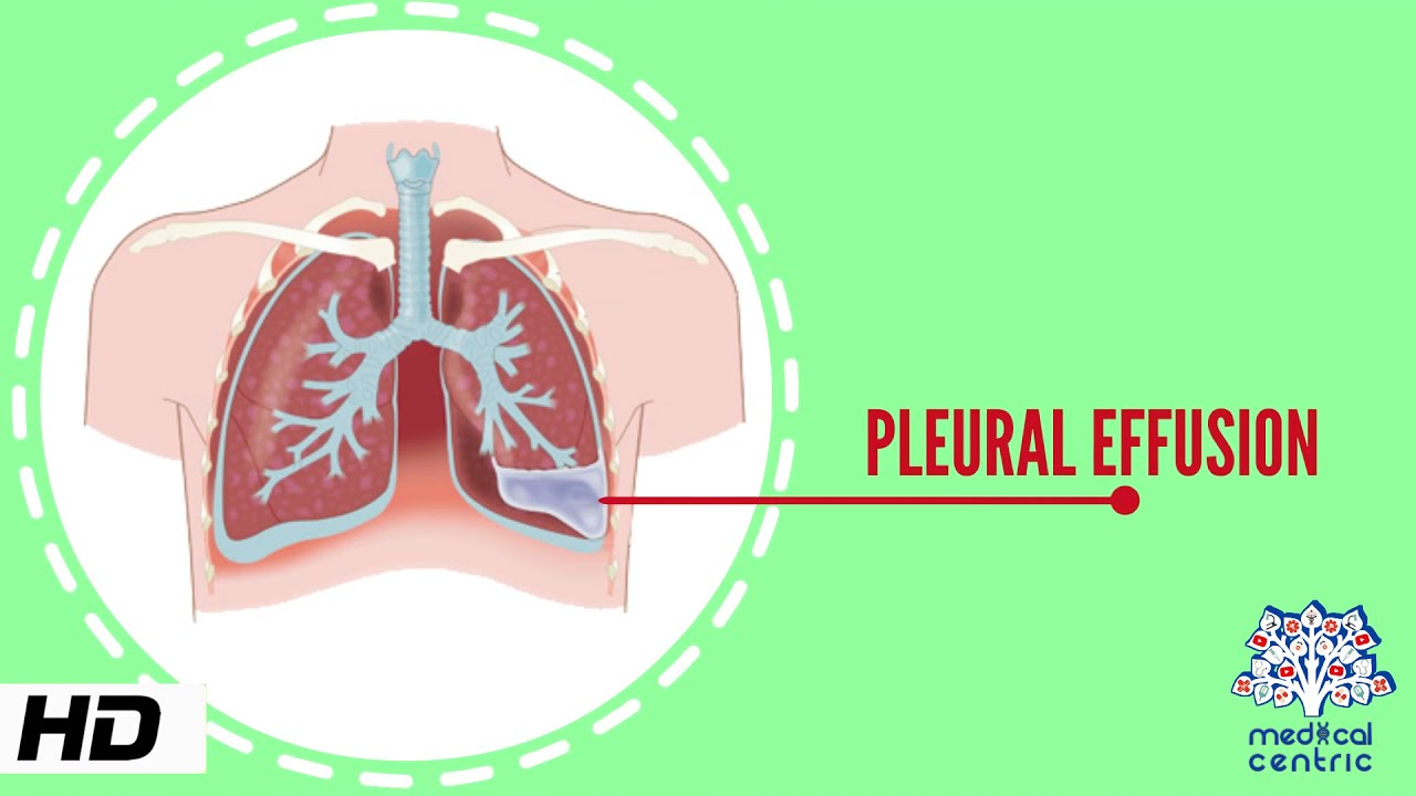
▫️BENIGN PLEURAL EFFUSION
▫️BENIGN PLEURAL THICKENING
▫️ASBESTOSIS: usually lower lobe predominant lung fibrosis
▫️MALIGNANT MESOTHELIOMA: a primary aggressive tumour of the pleura
👨🏽💻In this case the patient was also a smoker and by assessing the lung apices you will find a mass on the left. A more subtle finding is of mediastinal node enlargement with loss of the AP window corresponding with a node on the subsequent CT and PET-CT. Lung biopsy confirmed a primary lung cancer
👨🏽💻Remember to always be systematic: don’t forget the lung apices and mediastinal contour
🔻@theradiologistpage
———
✅Patient consent obtained
#theradiologist #radiology #radiologist #physician #physicianassistant #medicine #medstudent #medicalstudent #medschool #medicalschool #radtech #xray #medical #radiography #radiologystudent #doctor
Повторяем попытку...
Доступные форматы для скачивания:
Скачать видео
-
Информация по загрузке: