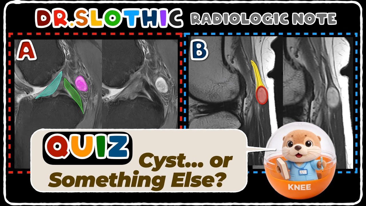
📌 Knee Synovial Recesses vs Schwannoma
Автор: MSK MRI Jee Eun Lee
Загружено: 2026-01-22
Просмотров: 252
Описание:
[Full Case Images]
📍 Watch Related Shorts
👉📌 Posterior Capsular Recess vs PCL Ganglion — Ultra Short Guide
• 📌 Posterior Capsular Recess vs PCL Ganglio...
👉📌 Key Imaging Features Suggesting Neurogenic Origin (PNSTs)
• 📌 Key Imaging Features Suggesting Neurogen...
✅ More structured MSK MRI guidance is available in my book,
Visualizing MSK Radiology: A Practical Guide to Radiology Mastery on Amazon.
https://www.amazon.com/dp/B0DJGMHMFS
📌 Knee Synovial Recesses vs Schwannoma
Central synovial recess
Anterior to the distal femur, behind the patella
Common site of anterior joint effusion
Hoffa-related recesses
Suprahoffatic: superior Hoffa’s fat pad → anterior synovitis
Infrahoffatic: anterior to ligamentum mucosum → cyclops vs synovitis pitfall
Posterior capsular recess (behind PCL)
Posterior extension of the medial femorotibial compartment
Frequently mimics a cystic lesion when distended
Parameniscal recess
Along superior/inferior meniscal margins
More prominent laterally; related to meniscal tears/cysts
Sub-popliteus recess
Between popliteus tendon and lateral meniscus
May communicate with the proximal tibiofibular joint
Posterior femoral (subgastrocnemius) recesses
Behind femoral condyles, deep to gastrocnemius
Common site of posterior effusion
Anterior tibial recess
Anterior to proximal tibia
Can simulate pathology if asymmetric
Posteromedial recesses (ramp-related)
Between medial femoral condyle, posterior horn of medial meniscus, and ramp capsule
Key in ramp lesion evaluation (ACL-deficient knee)
One Key Anatomic Insight
Medial meniscus: short, taut coronary ligament → less mobility
Lateral meniscus: redundant coronary ligament → greater mobility
→ Explains tear patterns and fluid tracking differences.
#KneeMRI #MSKRadiology #RadiologyEducation #SynovialRecess #KneeAnatomy #DiagnosticPitfalls #MRIInterpretation #OrthopedicImaging #RadiologyTeaching #DrSlothic
📌 Key Imaging Features Suggesting Neurogenic Origin (PNSTs)
1) Location Along Major Nerves
Masses located along the typical course of major nerves
(e.g., median nerve, sciatic nerve, tibial nerve)
should immediately raise suspicion for a neurogenic tumor.
2) Entering and Exiting Nerve
One of the strongest clues:
Visualization of a tubular nerve entering and exiting the mass.
This finding is considered pathognomonic for Peripheral Nerve Sheath Tumors (PNSTs).
3) Fusiform Shape
Lesions that are fusiform (spindle-shaped)
—elongated along the nerve’s axis—
are characteristic of neurogenic neoplasms and rare in soft tissue sarcomas.
4) Split-Fat Sign
A rim of fat surrounding the mass
→ known as the split-fat sign.
This suggests the tumor originated in the intermuscular fat plane
near the neurovascular bundle.
Best visualized on T1-weighted MRI.
Common in benign PNSTs.
5) Muscle Abnormalities
Changes in muscles supplied by the affected nerve can reinforce the diagnosis:
Fatty atrophy or decreased muscle bulk → best seen on T1
Edematous muscle changes from early denervation → best seen on T2
Always compare with the contralateral side for subtle cases.
Повторяем попытку...
Доступные форматы для скачивания:
Скачать видео
-
Информация по загрузке: