Muscle Histology Diagrams | How to Draw Skeletal, Cardiac & Smooth Muscle (TS & LS)
Автор: VB Anatomy
Загружено: 2026-02-03
Просмотров: 27
Описание:
📌 Muscle Histology Drawing Made Simple (For Beginners)
In this video, I guide you step-by-step through how to draw and label muscle histology diagrams, a topic that is very commonly asked in practical exams.
✅ Perfect for students who struggle with diagram corrections or microscopy slides.
✅ Slides Covered in This Video
1️⃣ Skeletal Muscle
Transverse section (TS) diagram
Longitudinal section (LS) diagram
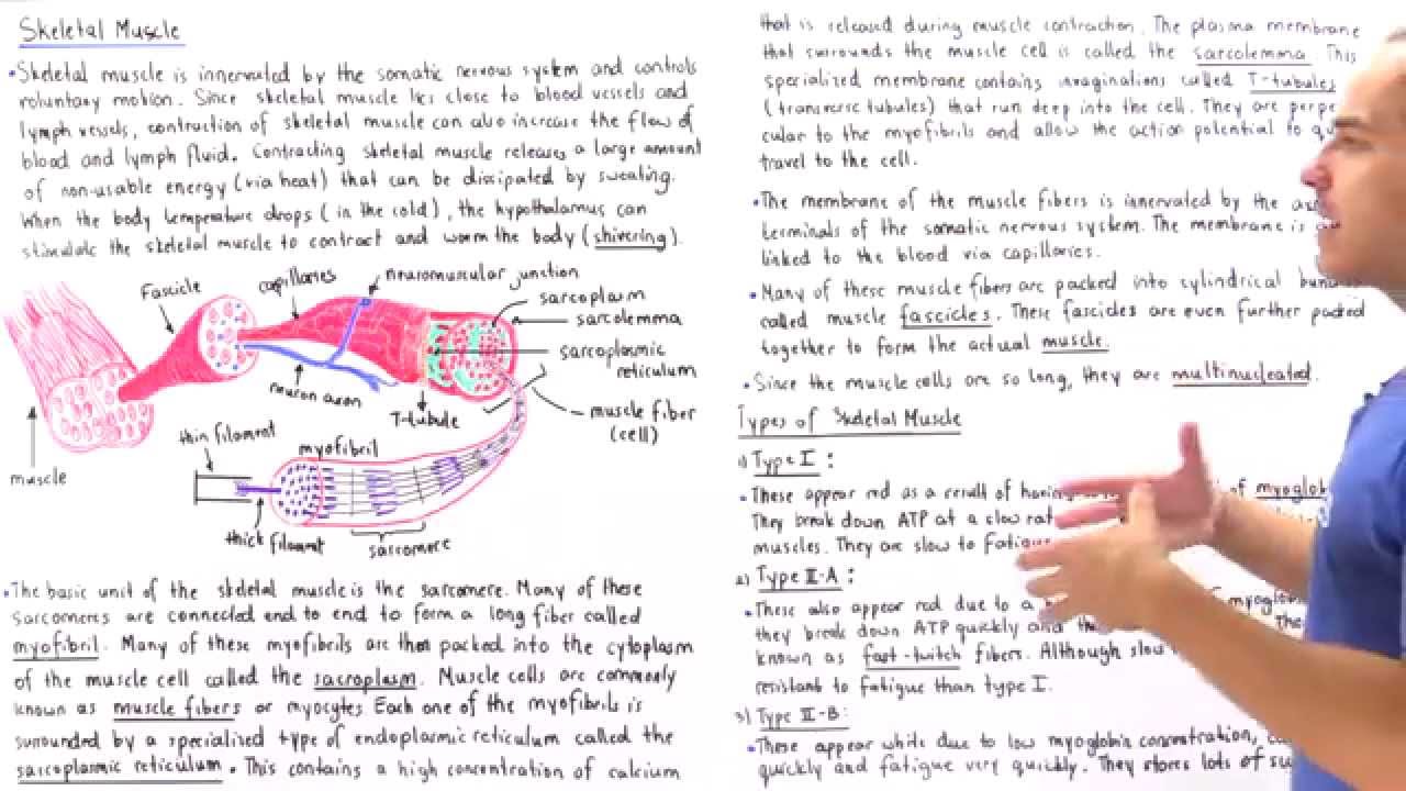
Peripheral flattened nuclei explained
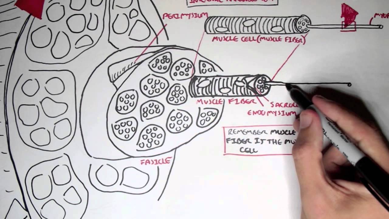
Endomysium, Perimysium & Epimysium concept
Myofibrils seen as tiny dots in TS
Sarcomere drawing: relaxed vs contracted
2️⃣ Cardiac Muscle
Branching fibers and syncytium formation
Intercalated discs clearly shown
Rounded central nuclei
Rich capillary supply highlighted
Ready-made comparison table: Skeletal vs Cardiac muscle
3️⃣ Smooth Muscle
Spindle-shaped cells and connective tissue
Why nuclei appear in some sections but not others
Smooth muscle found in vessel walls
Comparison table: Skeletal vs Smooth muscle
🎯 Useful For:
1st Year MBBS Students
BDS Students
Nursing Students
Physiotherapy Students
Ayurveda & Homeopathy Students
📚 Learn Histology Diagrams the Easy Way
This video will help you confidently answer questions like:
✔ Draw and label TS of skeletal muscle
✔ Draw cardiac muscle with intercalated discs
✔ Draw smooth muscle layers in vessels
🔔 Subscribe to VB Anatomy for more Sketch & Recall histology videos!
REFERENCE VIDEOS:
1 - Thyroid Gland – Histology Diagram
• Diagram of Histology of thyroid gland: A s...
2 - Transitional Epithelium
• Diagram of Transitional epithelium: A step...
3 - Microscopic Structure of Cerebellum (How to Draw)
• Diagram of Histology of Cerebellum: A step...
4 - Microscopic Structure of Bone – LS & TS (How to Draw)
• Mastering the drawing of Microscopic Bone ...
5 - Microscopic Structure of Salivary Gland (How to Draw)
• How to Draw the Microscopic Structure of S...
6 - Microscopic Structure of Retina (How to Draw)
• Microscopic structure of Retina
7 - Microscopic Structure of Cartilage (How to Draw)
• Cartilage Histology Made Easy | How to Dra...
8 - Connective Tissue Diagrams (Journal Practice)
• How to Draw Connective Tissue Histology Sl...
0:00 Introduction – Muscle Histology Diagrams Made Easy
0:10 Microscopic Structure of Muscle Tissue
0:30 Skeletal Muscle – Location and Examples
0:54 Draw & Label TS of Skeletal Muscle – Epimysium, Perimysium, Endomysium
2:25 Zoomed Diagram of TS Skeletal Muscle – Myofibrils Inside Muscle Fibre
3:55 Draw & Label LS of Skeletal Muscle – Striations and Peripheral Nuclei
5:24 Sarcomere Diagram – Relaxed vs Contracted (Most Asked Concept)
7:32 Draw & Label Cardiac Muscle – Intercalated Discs and Branching Fibres
9:58 Table: Differences Between Skeletal and Cardiac Muscle (Exam Comparison)
10:11 Draw & Label Smooth Muscle LS – Spindle Cells and Central Nuclei
11:30 Smooth Muscle TS – Why Nuclei Appear in Some Sections
13:10 Table: Differences Between Skeletal and Smooth Muscle (High-Yield)
Повторяем попытку...
Доступные форматы для скачивания:
Скачать видео
-
Информация по загрузке: